陶可医生的科普号
- 精选 2014年美国骨科医师协会AAOS官方杂志:股骨头坏死:评估和治疗
股骨头坏死:评估和治疗译者:陶可(北京大学人民医院骨关节科)摘要:股骨头坏死可能导致髋关节逐渐破坏。虽然股骨头坏死的病因尚未明确,但危险因素包括皮质类固醇的使用、饮酒、创伤和凝血(功能)异常。病变的大小和位置是疾病进展的预后因素,最好通过 MRI 进行评估。使用药物制剂和生物物理方法治疗股骨头坏死的疗效需要进一步研究。手术治疗基于患者因素和病变特征。对于没有头部塌陷的年轻患者,可以尝试通过使用血管化骨移植物、无血管移植物、骨形态发生蛋白、干细胞或上述方法的组合或旋转截骨术进行髓心减压来保护股骨头。最佳治疗方式尚未确定。当股骨头塌陷时,关节置换术是首选。文献出处:Charalampos G Zalavras, Jay R Lieberman. Osteonecrosis of the femoral head: evaluation and treatment. J Am Acad Orthop Surg. 2014 Jul;22(7):455-64.doi: 10.5435/JAAOS-22-07-455.Osteonecrosis of the femoral head: evaluation and treatmentAbstractOsteonecrosis of the femoral head may lead to progressive destruction of the hip joint. Although the etiology of osteonecrosis has not been definitely delineated, risk factors include corticosteroid use, alcohol consumption, trauma, and coagulation abnormalities. Size and location of the lesion are prognostic factors for disease progression and are best assessed by MRI. The efficacy of medical management of osteonecrosis with pharmacologic agents and biophysical modalities requires further investigation. Surgical management is based on patient factors and lesion characteristics. Preservation of the femoral head may be attempted in younger patients without head collapse by core decompression with vascularized bone grafts, avascular grafts, bone morphogenetic proteins, stem cells, or combinations of the above or rotational osteotomies. The optimal treatment modality has not been identified. When the femoral head is collapsed, arthroplasty is the preferred option.Lateral radiograph of the left hip (A) demonstrating presence of the crescent sign (arrows) and AP radiograph (B) demonstrating mild flattening of the femoral head (arrow).左侧髋关节 X线片(A)显示存在新月征(箭头),前后AP 位X 线片(B)显示股骨头轻度扁平(箭头)。Coronal T1-weighted magnetic resonance image of the right hip demonstrating a single-density line (arrow) of low signal intensity that delineates the area of necrotic bone.右髋的冠状 T1 加权核磁共振图像显示低信号强度的单密度线(箭头),描绘了坏死骨的区域。Modified Kerboul method. The arc of the necrotic portion of the femoral head on both midcoronal (A) and midsagittal (B) magnetic resonance images is measured, and the sum of the two angles is then calculated. (Reproduced with permission from Ha YC, Jung WH, Kim JR, Seong NH, Kim SY, Koo KH: Prediction of collapse in femoral head osteonecrosis: A modified Kerboul method with use of magnetic resonance images. J Bone Joint Surg Am 2006;88[suppl 3]:35-40.).改进的 Kerboul 方法。测量股骨头坏死部分在冠状面 (A) 和矢状面 (B) 磁共振图像上的弧度,然后计算两个角度的总和。(经 Ha YC、Jung WH、Kim JR、Seong NH、Kim SY、Koo KH 许可转载:股骨头坏死塌陷的预测:使用磁共振图像的改良 Kerboul 方法。J Bone Joint Surg Am 2006;88 [增刊 3]:35-40。)。Core decompression for a small osteonecrotic lesion seen on a coronal T1-weighted magnetic resonance image of the right hip (A). AP fluoroscopic images of the right hip show directing and centering the guidewire at the lesion (B), drilling over the guidewire to create a core tract (C), and removal of the necrotic bone with a burr (D). The femoral head is bone grafted with concentrated stem cells harvested from the iliac crest. The core tract is sealed with demineralized matrix.在右侧髋关节的冠状 T1 加权核磁共振图像上看到的一个小的股骨头坏死病变的髓心减压(A)。右髋关节 AP位 透视图像显示将导丝引导并居中在病变处 (B),在导丝上钻孔以创建髓心通道 (C),并用球形铰刀头去除坏死骨 (D)。股骨头(坏死区域)用从髂嵴采集的浓缩干细胞进行骨髓移植。髓心通道用脱矿质基质填充。Vascularized fibula graft. The fibular graft is inserted into the core tract and stabilized with a Kirschner wire (K). The peroneal veins and artery are anastomosed to the ascending branches of the lateral femoral circumflex artery (LFCA) and vein.血管化腓骨移植物。将腓骨移植物插入髓心通道并用克氏针 (K) 固定。腓静脉和动脉与旋股外侧动脉(LFCA)和静脉的升支吻合。
陶可 主治医师 北京大学人民医院 骨关节科690人已读 - 精选 2020年最新JBJS文献——非创伤性股骨头坏死:我们今天的立场(研究进展)?:5 年更新
非创伤性股骨头坏死:我们今天的立场(研究进展)?:5 年更新译者:陶可(北京大学人民医院骨关节科)摘要: 临床医生应高度警惕高危患者(使用皮质类固醇、过量饮酒、患有镰状细胞病等),以便及早诊断出股骨头坏死。 非手术治疗方式在阻止(股骨头坏死)进展方面通常是无效的。因此,当人们试图保留天然(髋)关节时,非手术治疗在早期是不合适的,除非在极少数情况下,小尺寸、位于内侧的病变可能无需手术即可愈合。 早期病变应尝试保髋手术,以保留住股骨头。 基于细胞疗法的(髋)关节保留手术继续显示出有希望的结果,因此应被视为可能改善临床结果的辅助治疗方法。 在骨坏死的情况下全髋关节置换术的结果非常好,结果与潜在诊断为骨关节炎的患者的结果相似。文献出处:Michael A Mont, Hytham S Salem, Nicolas S Piuzzi, Stuart B Goodman, Lynne C Jones. Nontraumatic Osteonecrosis of the Femoral Head: Where Do We Stand Today?: A 5-Year Update. Review, J Bone Joint Surg Am. 2020 Jun 17;102(12):1084-1099.doi: 10.2106/JBJS.19.01271.Nontraumatic Osteonecrosis of the Femoral Head: Where Do We Stand Today?: A 5-Year UpdateAbstract Clinicians should exercise a high level of suspicion in at-risk patients (those who use corticosteroids, consume excessive alcohol, have sickle cell disease, etc.) in order to diagnose osteonecrosis of the femoral head in its earliest stage. Nonoperative treatment modalities have generally been ineffective at halting progression. Thus, nonoperative treatment is not appropriate in early stages when one is attempting to preserve the native joint, except potentially on rare occasions for small-sized, medially located lesions, which may heal without surgery. Joint-preserving procedures should be attempted in early-stage lesions to save the femoral head. Cell-based augmentation of joint-preserving procedures continues to show promising results, and thus should be considered as an ancillary treatment method that may improve clinical outcomes. The outcomes of total hip arthroplasty in the setting of osteonecrosis are excellent, with results similar to those in patients who have an underlying diagnosis of osteoarthritis.Figs. 1-A and 1-B Small-diameter CD for ONFH. A trocar is introduced into the necrotic lesion using light mallet blows (Fig. 1-A) under fluoroscopic guidance (Fig. 1-B).图1 用于股骨头坏死 ONFH 的 1-A 和 1-B 小直径髓心减压CD术。在透视引导下(图 1-B)使用轻槌敲击(图 1-A)将套管针引入坏死病变区域。Fig. 2 Aspiration of bone marrow from the iliac crest for subsequent processing and implantation following femoral head CD.图 2 从髂嵴抽吸骨髓,用于股骨头髓心减压CD术后的后续处理和植入。Fig. 3 The lightbulb technique—creation of a cortical window at the femoral head-neck junction for evacuation of necrotic tissue and replacement with a bone graft.图 3 灯泡技术——在股骨头颈交界处创建一个皮质窗口,用于清除坏死组织并用骨移植物替代。
陶可 主治医师 北京大学人民医院 骨关节科1509人已读 - 精选 股骨头坏死:病因、诊断及治疗方式:2019英国医学杂志(BMJ)(医学生及低年资住院医师培训之)综述
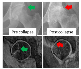
股骨头坏死:病因、诊断及治疗方式:2019英国医学杂志(BMJ)(医学生及低年资住院医师培训之)综述作者:JonathanNLamb,ColinHolton,PhilipO'Connor,PeterVGiannoudis作者单位:LeedsInstituteofRheumaticandMusculoskeletalMedicine,SchoolofMedicine,UniversityofLeeds,Leeds,UK.译者:陶可(北京大学人民医院骨关节科)股骨头坏死你需要了解什么?•股骨头坏死(AVNFH)的常见危险因素包括:酗酒、使用类固醇激素、化疗和免疫抑制剂药物以及镰状细胞性贫血。•如果患者髋部疼痛持续超过6周且X线片正常,请考虑对髋部进行核磁共振MRI扫描,并转诊至骨科(保髋治疗)团队。•早期治疗可使髋关节7年后存活率提高至88%。Fig1Demonstrationofhiprotationtoelicithippainwiththepatientsitting(A)andsupine(B,C,D).图1 演示通过患者坐位(A)和仰卧位(B、C、D)引起髋部疼痛的髋关节旋转活动(髋关节查体之内外旋转活动)。Fig2Typicalchangesseenonplainradiograph(top)andMRI(bottom)ofthehipinearlyandlateAVNFH.TheappearanceofearlyAVNFHisnotapparentonplainradiographbutisvisibleonMRI.图2 早期和晚期股骨头坏死(AVNFH)髋关节平片(上)和MRI(下)中看到的典型变化。早期股骨头坏死(AVNFH)的在平片上表现不明显,但在MRI上可见明显信号的改变。Fig3ProposedpathwayformanagingAVNFHinaprimarycaresetting.图3 在基层医疗机构中管理股骨头坏死(AVNFH)的建议诊治流程。 典型病例:一名36岁的女性向她的全科医生报告,有左侧腹股沟疼痛放射到膝盖的病史。疼痛很严重,走路时更严重,并伴有跛行。一年后,患者再次去看全科医生,尽管进行了镇痛,但疼痛仍持续存在。髋关节和膝关节的平片显示髋关节间隙轻微变窄,没有其他特征,她被转诊到二级骨科诊所。髋关节磁共振成像(MRI)扫描显示股骨头缺血性坏死(AVNFH)伴塌陷的典型特征。什么是股骨头缺血性坏死?股骨头坏死(AVNFH)由于微循环异常而导致软骨下骨结构完整性丧失。潜在的发病机制尚不清楚;风险因素可能会以某种方式影响微循环,但这尚未得到研究证实。共同的终点是微循环异常和坏死。软骨下骨随后塌陷,导致进行性、继发性髋关节骨关节炎。在英国,平均发病年龄为58.3岁,每10万名患者中有2人患病。平均而言,股骨头坏死(AVNFH)比典型骨关节炎发生得更早。它在男性中更为常见,发病率最高的是25至44岁的男性和55至75.3岁的女性。在英国,它是50.4岁以下人群全髋关节置换术的第三大常见适应症。以下因素与股骨头坏死(AVNFH)风险增加相关:•血液甘油三酯、总胆固醇、低密度脂蛋白胆固醇和非高密度脂蛋白胆固醇水平高;•男性;•城市居民;•股骨头坏死(AVNFH)家族史;•大量吸烟;•滥用酒精;•超重;•凝血病;•血管病变;•艾滋病病毒;•大量接触类固醇激素、化疗和免疫抑制药物。类固醇激素已被证明会使骨坏死(非部位特异性)的几率增加3倍,而免疫抑制剂则增加6倍。Zhao报告称,服用皮质类固醇激素的患者发生股骨头坏死(AVNFH)的几率高出35倍,患有“酗酒”状态的患者则高出6倍。为什么股骨头坏死(AVNFH)会漏诊呢?股骨头坏死(AVNFH)很少见。患有这种疾病的患者可能同时患有慢性风湿病和血液病。这可能会导致诊断的不确定性,特别是考虑到在这些情况下使用化疗、免疫调节剂和类固醇激素时,这些都是股骨头坏死(AVNFH)的危险因素。查体可以帮助识别可能引起疼痛的解剖结构,因为髋关节疼痛可能源自髋关节和非髋关节的多个部位。临床表现可能会被错过,因为由于时间和空间的限制,在基层医疗机构中准确确定单纯由于髋关节运动时造成的腹股沟疼痛可能具有挑战性(比较困难)。股骨头坏死(AVNFH)早期阶段的正常X线片可能会错误地让人放心并延迟适当的转诊。如果X线片呈阴性并且患者继续抱怨髋关节疼痛,医生可能会诊断为非特异性髋关节疼痛(考虑到肌肉骨骼的原因)并建议患者接受物理治疗。在新发病例中,18.75%只能通过MRI进行诊断,并且在普通X线片上很容易被漏诊。只有MRI扫描才具有诊断意义。为什么及早确诊股骨头坏死(AVNFH)很重要?早期诊断和转诊至关重要,因为骨质破坏通常发生在发病后2年内,因此不可能进行保留髋关节的治疗干预(保髋治疗在股骨头坏死发病2年内)。股骨头坏死(AVNFH)的早期发现使多学科团队有时间改变可能引发股骨头坏死(AVNFH)发作的治疗方法。股骨头髓心减压术可降低中短期内进一步手术的需要,但仅适用于疾病的最早阶段。一旦患者进展为继发性髋关节骨关节炎,关节置换通常是不可避免的。然而,考虑到股骨头坏死(AVNFH)患者年龄较小,翻修手术和相关发病率的终生风险很大。如何诊断股骨头坏死(AVNFH)?股骨头坏死(AVNFH)诊断从仔细询问病史和检查开始,以确定髋关节疼痛的来源。最终需要MRI来诊断股骨头坏死(AVNFH),并且还可以诊断髋关节疼痛的其他原因。仔细的询问病史病史显示疼痛持续超过6周,通常位于腹股沟和大腿,负重和运动时疼痛更严重。通常没有外伤史。询问危险因素,如果患者有任何“危险信号”,请进行髋关节MRI检查。股骨头坏死(AVNFH)通常是双侧的,双侧股骨头坏死(AVNFH)的风险通常是在单侧确诊后的2年内。框1:需要转介或进一步评估的危险信号:•髋关节X线检查正常,髋关节疼痛超过6周;•患有髋关节疼痛和危险因素的患者,包括:o既往单侧股骨头坏死(AVNFH),o酗酒,o大量接受类固醇激素治疗,o免疫治疗,Ø化疗,o镰状细胞病和其他凝血病,Ø艾滋病毒,o新近妊娠。查体腹股沟、大腿和膝关节前侧疼痛的再现并伴有单独的大腿旋转不能诊断股骨头坏死(AVNFH),但有助于区分髋关节疼痛与脊柱和膝关节疼痛。这可以在患者坐位或仰卧时进行(图1)。影像学检查早期股骨头坏死(AVNFH)在X线片上并不明显。如果患者持续感到疼痛,则需要进一步检查和转诊。股骨头坏死(AVNFH)通过髋关节MRI进行诊断,当与临床症状密切相关时,还可以诊断各种可治疗的髋关节疼痛(例如风湿病、肌腱疾病和骨病)(图2)。仅当有其他原因或高度怀疑风湿病或感染时,才应考虑进行其他检查,例如血液检查。转诊如果患者的髋关节MRI显示有股骨头坏死(AVNFH)改变时,请就诊于骨科医生(图3)。在二级医疗机构就诊时,股骨头坏死(AVNFH)诊断应与开具类固醇激素、化疗和免疫治疗原发病的任何治疗团队共享。药物和手术治疗取决于患者的特征和股骨头坏死(AVNFH)的阶段。使用前列环素类似物和双膦酸盐对塌陷前股骨头坏死(AVNFH),可以减轻症状并防止关节形合度破坏,但其疗效目前尚不清楚。手术治疗仍存在争议,但大多数塌陷前股骨头坏死(AVNFH)患者均接受髓心减压手术,并辅以或不辅以药物治疗,以减轻疼痛,并有可能在长达7年的时间里避免88%的患者进行全髋关节置换术治疗。术后恢复包括12个月的非负重康复锻炼,并在8周后逐渐恢复工作和驾驶。通常在术后12个月即可感受到完全的治疗益处。专业的三级医疗机构可以提供新的治疗方法,例如骨移植和截骨术,以分别促进血管再生和减轻受损髋关节表面的负荷。一旦发生塌陷,全髋关节置换术可以为患者提供快速、可靠的疼痛缓解和功能改善,但与未来有髋关节翻修的风险,特别是对于年轻患者。 AvascularnecrosisofthehipWhatyouneedtoknow•CommonriskfactorsforAVNFHarealcoholism,useofsteroids,chemotherapyandimmunosuppressantmedication,andsicklecellanaemia.•ConsiderMRIscanofthehipandreferraltoanorthopaedicteamifapatienthasapainfulhipforlongerthansixweekswithnormalradiographs.•Earlytreatmentimprovesthechancesofhipsurvivalbyupto88%atsevenyears. A36yearoldwomanpresentstoherGPwithahistoryofleftgroinpainradiatingtotheknee.Thepainissevere,worseonwalking,andassociatedwithalimp.ThepatientrevisitstheGPayearlaterwithpersistentpaindespiteanalgesia.Plainradiographsofthehipandkneeshowslightnarrowingofthehipjointspacewithnootherfeaturesandsheisreferredtoasecondarycareorthopaedicclinic.Amagneticresonanceimaging(MRI)scanofthehipshowsclassicfeaturesofavascularnecrosisofthefemoralhead(AVNFH)withcollapse.Whatisavascularnecrosisofthefemoralhead?Osteonecrosisofthefemoralhead(AVNFH)causeslossofintegrityofsubchondralbonestructureduetoabnormalmicrocirculation.Theunderlyingpathogenesisisunclear1;riskfactorsarelikelytoaffectmicrocirculationinsomewaybutthishasnotbeenconfirmedbyresearch.Thecommonendpointisabnormalmicrocirculationandnecrosis.Subchondralbonesubsequentlycollapses,whichleadstoprogressivesecondaryarthritis.MeanageofpresentationintheUKis58.3years,withaprevalenceoftwoper100000patients.2Onaverage,AVNFHoccursearlierinlifethantypicalosteoarthritis.Itismorecommoninmenandthehighestprevalenceisinmenaged25to44andwomenaged55to75.3IntheUKitisthethirdmostcommonindicationfortotalhipreplacementinpeopleunder50.4ThefollowingfactorsareassociatedwithanincreasedriskofAVNFH35:•Highlevelsofbloodtriglycerides,totalcholesterol,lowdensitylipoproteincholesterol,andnon-highdensitylipoproteincholesterol•Malesex•Urbanresidence•FamilyhistoryofAVNFH•Heavysmoking•Alcoholabuse•Overweight•Coagulopathies•Vasculopathies•HIV•Highexposuretosteroids,chemotherapy,andimmunosuppressantmedication.Steroidshavebeenshowntoincreaseoddsofosteonecrosis(non-sitespecific)byafactorofthreeandimmunosuppressantsbyafactorofsix.ZhaoreportedthattheoddsofAVNFHwere35timesgreaterinpatientstakingcorticosteroidsandsixtimesgreaterinpatientswith“alcoholism”status.3Whyisitmissed?AVNFHisrare.Patientswiththeconditioncanhavecoexistingchronicrheumaticandhaematologicalproblems.Thismayleadtodiagnosticuncertainty,particularlygiventheuseofchemotherapy,immunomodulatoryagents,andsteroidsintheseconditions,whichareallriskfactorsforAVNFH.Aphysicalexaminationcanhelpidentifytheanatomicalstructuresthatmightbecausingthepain,sincehippaincanoriginatefrommultiplehipandnon-hipareas.Presentationsmaybemissedbecauseaccuratereproductionofgroinpainonisolatedhipmovementscanbechallengingtoelicitinaprimarycaresettingduetotimeandspaceconstraints.NormalplainradiographsintheearlystagesofAVNFHcanbefalselyreassuringanddelayappropriatereferral.Iftheplainradiographisnegativeandthepatientcontinuestocomplainofhippain,thedoctormaygiveadiagnosisofnon-specifichippain(giventhatmusculoskeletalpresentationsarecommoninprimarycare)andsendthepatientforphysiotherapy.Ofnewpresentations,18.75%arediagnosableonlywithMRIandareeasilymissedonnormalplainradiographs.3OnlytheMRIscanisdiagnostic.Whydoesitmatter?Earlydiagnosisandreferralareessentialsincebonedestructionnormallyoccurswithintwoyearsofdiseaseonset,makingjointpreservinginterventionimpossible.6EarlyidentificationofAVNFHgivesthemultidisciplinaryteamtimetochangemedicaltreatmentswhichmightbeprovokingonsetofAVNFH.Surgicaldecompressionofthefemoralheadreducestheneedforfurthersurgeryintheshorttomediumtermbutisonlysuitablefortheearlieststagesofdisease.5Oncepatientshaveprogressedtosecondaryhiparthritis,jointreplacementisusuallyinevitable.However,giventheyoungerageofpatientswithAVNFH,thelifetimeriskofrevisionsurgeryandassociatedmorbidityisgreat.HowisAVNFHdiagnosed?AVNFHdiagnosisstartswithacarefulhistoryandexaminationtodeterminethatthehipisthesourceofpain.UltimatelyanMRIisrequiredtodiagnoseAVNFHandmayalsodiagnoseothercausesofhippain.AcarefulhistoryAhistoryshowingpainlastinglongerthansixweeks,typicallylocatedinthegroinandthighandwhichisworseonweightbearingandmovementiskey.6Usuallythereisnohistoryoftrauma.AskaboutriskfactorsandreferforMRIofthehipifthepatienthasany“redflags”(box1).AVNFHisoftenbilateralandtheriskofbilateralAVNFHishighestwithintwoyearsofunilateraldiagnosis.6Box1:Redflagsrequiringreferralorfurtherassessment•Hippainformorethansixweekswithnormalhipradiograph•PatientspresentingwithhippainandriskfactorsincludingopreviousunilateralAVNFHoalcoholexcessohighexposuretosteroidtherapyoimmunologictherapyochemotherapyosicklecelldiseaseandothercoagulopathiesoHIVorecentpregnancyExaminationReproductionofpaininthegroin,thigh,andanterioraspectofkneewithisolatedthighrotationwillnotdiagnoseAVNFH,butwillhelptodifferentiatehippainfrompainoriginatingfromthespineandknee.Thiscanbeperformedwiththepatientsittingorsupine(fig1).RadiologicaltestsEarlyAVNFHisnotapparentonplainradiographs.Ifthepatientcontinuestobeinpain,furtherinvestigationandreferraliswarranted.AVNFHisdiagnosedonMRIofthehips,7whichmayalsodiagnoseabreadthoftreatablehippain(suchasrheumatologicaldisease,musculotendinousdisease,andbonydisease)whencarefullycorrelatedwithclinicalsymptoms8(fig2).Otherinvestigations,suchasbloodtests,shouldonlybeconsideredifindicatedforotherreasonsorifthereisahighsuspicionofrheumatologicaldiseaseorinfection.ReferralIfthepatienthassignsofAVNFHonMRIofthehip,refertoanorthopaedicsurgeonforconsultation(fig3).Insecondarycare,AVNFHdiagnosisshouldbesharedwithanycareteamsinvolvedintheadministrationofsteroids,chemotherapy,andimmunologictherapy.MedicalandsurgicaltreatmentdependonthepatientcharacteristicsandstageofAVNFH.Medicaltreatmentofpre-collapsediseasewithprostacyclinanaloguesandbisphosphonatesmayreducesymptomsandpreventlossofjointcongruitybuttheirefficacyisnotcurrentlywelldefined.6Surgically,treatmentremainscontroversial,butmostpatientswithpre-collapseAVNFHareofferedcoredecompressionsurgerywithorwithoutadjunctivepharmacologicaltherapytoreducepainandpotentiallypreventtheneedfortotalhipreplacementin88%ofpatientsforuptosevenyears.910Postoperativerecoveryinvolvesaperiodofnon-weightbearingfor12monthsandgradualreturntoworkanddrivingat8weeks.Fullbenefitisusuallyfeltat12monthsaftersurgery.Specialisttertiarycentresmayoffernoveltreatmentssuchasbonegraftingandosteotomiestoencouragevascularregrowthandunloaddamagedhiparticularsurface,respectively.Oncecollapsehasoccurred,totalhipreplacementcangivepatientsrapid,reliablepainreliefandimprovedfunctionbutisassociatedwiththeriskoffuturerevision,particularlyinyoungerpatients.Afulldescriptionofalltheoptionsisbeyondthescopeofthisarticleandpatientsshoulddiscussallavailableoptionswiththeirsurgeontoenableinformedshareddecisionmaking.文献出处:JonathanNLamb,ColinHolton,PhilipO'Connor,PeterVGiannoudis.Avascularnecrosisofthehip.BMJ.2019May30;365:l2178.doi:10.1136/bmj.l2178.
 陶可 主治医师 北京大学人民医院 骨关节科514人已读
 - 精选 股骨头坏死分类:哪些患者(需要且)应该手术?2019年(日本JIC分型:保头/保髋手术适应证)
股骨头坏死分类:哪些患者(需要且)应该手术?2019年(日本JIC分型:保头/保髋手术适应证)作者:YKuroda,TTanaka,TMiyagawa,TKawai,KGoto,STanaka,SMatsuda,HAkiyama作者单位:DepartmentofOrthopaedicSurgery,GraduateSchoolofMedicine,KyotoUniversity,Kyoto,Japan.译者:陶可(北京大学人民医院骨关节科)摘要目的:使用简单的分类方法,我们旨在估计股骨头坏死(ONFH)导致的塌陷率,以制定保留髋关节的手术治疗指南。方法:我们回顾性分析诊断为ONFH的310名患者(141名男性,169名女性;平均年龄45.5岁(标准差14.9;15至86))的505侧髋关节,并使用日本调查委员会(JIC)分类对其进行分类。JIC系统根据承重表面上骨坏死病变的位置和大小(A、B、C1和C2型)和ONFH阶段包括四种可视化类型。使用Kaplan-Meier生存分析计算由ONFH引起的塌陷率,以影像学塌陷/关节置换术为终点。结果:双侧390髋,单侧115髋。按JIC分型,A型21髋,B型34髋,C1型173髋,C2型277髋。初步诊断时,238/505髋(47.0%)已经塌陷。此外,对212例塌陷前髋关节的累积存活率进行分析,发现2年和5年塌陷率A、B、C1和C2型分别为0%和0%、7.9%和7.9%、23.2%和36.6%、57.8%和84.8%。结论:A型ONFH无需进一步治疗,但C2型塌陷前ONFH需要立即进行保髋手术治疗。考虑到高塌陷率,我们的研究结果证明了对C2型ONFH无症状患者进行早期诊断和干预的重要性。图1 日本骨坏死调查委员会分类系统(JIC)分型图示表1 患者人口统计数据具有统计学意义†Kruskal–Wallis检验‡对数秩检验Kaplan–Meiersurvivalcurvesofprecollapsecases.a)Thecumulativefive-yearsurvivalratesindicatethatthecollapserateofprecollapseosteonecrosisofthefemoralhead(ONFH)casesis0%to84.8%,intheorderofsmallertolargerlesionsizes.TypeC2progressedquickly,with37%atoneyearand58%attwoyearsreachingtheendpoint.b)CollapserateofprecollapseONFHcasesaccordingtosex;therewerenodifferencesintermsoftimetocollapse(p=0.453,log-ranktest).c)CollapserateofprecollapseONFHcasesaccordingtolaterality;therewerenodifferencesintermsoftimetocollapse(p=0.580,log-ranktest).d)CollapserateofprecollapseONFHcasesaccordingtosteroiduse;therewerenodifferencesintermsoftimetocollapse(p=0.961,log-ranktest).图2 塌陷前病例的Kaplan-Meier生存曲线。a)累积五年生存率表明,股骨头塌陷前骨坏死(ONFH)病例的塌陷率为0%至84.8%,按病变大小从小到大的顺序排列。C2型进展迅速,一年达到终点的比例为37%,两年时达到终点的比例为58%。b)按性别划分的塌陷前ONFH病例的塌陷率;在塌陷时间方面没有差异(p=0.453,对数秩检验)。c)根据外侧边缘的塌陷前ONFH病例的塌陷率;在塌陷时间方面没有差异(p=0.580,对数秩检验)。d)根据类固醇使用情况,塌陷前ONFH病例的塌陷率;在塌陷时间方面没有差异(p=0.961,对数秩检验)。 Five-yearcollapseratesandhazardratios(HRs)ofeachdiseasetypeasevaluatedbytheCoxregressionmodel.AhighercollapserateandanincreaseinHRforcollapseofthefemoralheadcanbeseenastheosteonecroticlesionsizeincreases.图3 通过Cox回归模型评估的每种疾病类型的五年塌陷率和风险比(HR)。随着股骨头坏死病灶大小的增加,可以看出股骨头塌陷的塌陷率和HR增加。 表3 日本调查委员会分类系统评估的其他塌陷率报告 症状的发生率†通过Kaplan-Meier生存分析评估的塌陷率 讨论几十年来,无症状ONFH患者的最佳手术治疗方案一直是一个有争议的话题。考虑到较高塌陷率,一些研究人员建议早期诊断和干预以实现ONFH患者的髋关节保留,包括无症状的ONFH患者。相反,另一些作者建议仅对有症状的ONFH患者进行手术。由于缺乏可靠的塌陷率数据,这个问题被认为是有争议的跨越不同类型的ONFH。此外,目前世界范围内的ONFH分类系统很多,包括Ficat和Arlet、骨循环研究协会(ARCO)、Steinberg和JIC。在这些分类系统中,ARCO、Steinberg和JIC系统包括基于病变大小和位置的分类。特别是,JIC分类具有一些优点,例如基于涉及髋臼的ONFH病变的大小和位置进行分类head.该系统包括四种简单类型,具有较高的观察者内和观察者间可靠性。在之前的系统评价中,Sultan等报告说,JIC分类在保持简单性的同时显示出良好的预后价值。在日本,为了获得用于医疗支持的难治性疾病状态登记卡,需要每年报告JIC阶段和类型。年度报告必须由日本骨科协会认证的医师撰写,其数据每年都会更新。因此,JIC分类在日本被广泛使用。与以往研究的结果一致,本研究发现,随着承重表面的股骨头坏死病变增大,塌陷率增加。Kaplan-Meier生存分析是确定塌陷率和评估股骨头塌陷的实际时间间隔的最佳方法。在这项研究中,A型髋关节的塌陷率为0%,C2型的塌陷率为84.8%,为确定最佳治疗方法提供了有用的线索。A型ONFH的最低塌陷率表明对该类型ONFH的进一步治疗是不合理的。相比之下,C2型ONFH患者建议进行保留髋关节的手术治疗,因为保守治疗不太可能实现保髋效果。C1型患者的早期干预应根据手术是否会给患者带来负担来确定。我们认为微创治疗,例如髓心减压或基于髓心减压的再生治疗,适合C1型患者。在Mont等之前的系统评价中,根据ONFH的不同分类系统评估了总共664个髋关节,据报道,59%的病例进展为(髋痛)症状或塌陷。据报道,股骨头塌陷的可能性与较大的病变大小和外侧承重表面的定位相关。此外,之前的几项研究使用JIC分类系统报告了ONFH的塌陷率(表III),并且其他研究人员报告了C型ONFH的较小塌陷率(在71%和76%之间)。Hungerford和Jones报道,小于15%的股骨头(按体积)的小病灶不太可能进展,这表明进展取决于病灶大小。除了JIC分类外,Steinberg分类被广泛使用,因为它结合了分期和按百分比评估受影响的病灶大小(<15%、15%至30%和>30%)。Min等强调考虑到C2型ONFH的高塌陷率,塌陷的重要预后因素不仅包括坏死病变的大小,还包括坏死病变的范围和位置。Kerboul角也被用于评估病变大小,并包括在前后位和外侧X线片上测量的两个角度的相加。在本研究中,我们展示了另一个显著的发现:根据(股骨头坏死的)外侧位置,在初始诊断时的ONFH阶段方面存在显着差异。据我们所知,以前没有报告对双侧和单侧ONFH进行直接比较。双侧ONFH被认为比单侧ONFH更容易诊断,并且认为使用口服药物治疗类固醇引起的骨质疏松症在双侧ONFH比单侧ONFH可能性更高。Mont等报道,无症状ONFH通常见于有症状ONFH患者的对侧髋关节。据报道,双侧ONFH的发生率高达75%。此外,据报道,根据背景因素(类固醇、未使用类固醇),初始诊断时ONFH的分期存在差异。对于免疫学家和骨外科医生来说,ONFH已被认为是全身性皮质类固醇治疗的不良事件。使用类固醇的患者被广泛认为是ONFH潜在风险较高的人群。此外,我们推测使用类固醇的ONFH患者的发生率显着降低,因为与未使用类固醇的患者相比,他们更容易被诊断出来。本研究存在一些局限性。首先,JIC分类的观察者内部和观察者间可靠性没有根据先前研究报告的高可靠性进行评估。Nakamura等报告在JIC分类的观察者间可靠性方面有85%的一致性(加权kappa:0.71)和82%的观察者内部可靠性一致(加权kappa:0.78)。Takashima等报道,JIC观察者间的可靠性被证明是可接受的(加权kappa:0.72),其可靠性高于Steinberg和Kerboul分类(均p<0.001,Spearman等级相关系数)。其次,我们没有评估口服药物患者或接受手术治疗的患者的结局。我们的数据反映了亚洲截骨术的流行程度。第三,我们没有分析症状与股骨头塌陷之间的关系。塌陷或塌陷后再生的方式可能会影响与髋关节相关的症状。Bozic等报道,髓心减压后失败率增加超过四倍与ONFH相关的骨囊性变有关。此外,通过破骨细胞再吸收的骨吸收受到修复过程的抑制,并表明ONFH的硬化变化。最后,ONFH患者背景因素的流行在世界范围内各不相同。然而,患者背景因素和ONFH的分类存在国际差异,总结500个或更多ONFH的报道很少。我们相信我们的研究填补了这一空白。总之,使用JIC分类预测股骨头塌陷有助于选择塌陷前ONFH患者的治疗策略。近几十年来,见证了新疗法的发展,例如自体骨髓细胞移植、金属植入棒的使用、和生长因子疗法。我们的研究结果证明对具有较大骨坏死病变的塌陷前、无症状ONFH患者进行早期干预是合理的,尤其是在C2型ONFH患者中。研究结果表明,A型ONFH不需要进一步的手术干预。DiscussionForseveraldecades,theoptimalsurgicaltreatmentoptionforpatientswithasymptomaticONFHhasremainedacontroversialtopic.1-3,5,9,18,22Consideringthehighcollapserate,severalresearchershaverecommendedearlydiagnosisandinterventiontoachievejointpreservationinONFHpatients,includingasymptomaticONFHpatients.1-3,5,18Incontrast,someauthorshaverecommendedsurgeryonlyforsymptomaticONFHpatients.14,15,17ThisissueisconsideredcontentiousowingtothelackofreliabledataonthecollapserateacrossthedifferenttypesofONFH.Inaddition,therearecurrentlymanyclassificationsystemsforONFHworldwide,includingtheFicatandArlet,AssociationResearchCirculationOsseous(ARCO),Steinberg,andJIC.Oftheseclassificationsystems,theARCO,Steinberg,andJICsystemsincludeclassificationbasedonlesionsizeandlocation.1,2Inparticular,theJICclassificationhassomeadvantages,suchasclassificationbasedonthesizeandlocationofONFHlesionsinvolvingtheacetabularhead.5,10,20Thissystemcomprisesfoursimpletypesandhashighintraobserverandinterobserverreliabilities.8,10,23Inaprevioussystematicreview,Sultanetal8reportedthattheJICclassificationshowspromisingprognosticvaluewhilemaintainingsimplicity.InJapan,annualreportingoftheJICstageandtypeisnecessaryinordertoobtainanintractablediseasestatuscardformedicalsupport.AnnualreportshavetobewrittenbyaJapaneseOrthopaedicAssociation–certifiedphysician,anditsdataareupdatedeveryyear.21Therefore,theJICclassificationiswidelyusedinJapan.Consistentwiththefindingsofpreviousstudies,thepresentstudyfoundthatthecollapserateincreasesastheosteonecroticlesionontheweightbearingsurfacebecomeslarger.1-3,10-15,22Kaplan–Meiersurvivalanalysisistheoptimalmethodfordeterminingthecollapserateandevaluatingtheactualintervaloffemoralheadcollapse.10,15,16,24Inthisstudy,typeAhipshada0%collapserateandtypeC2hadan84.8%collapserate,providingusefulcluesfordeterminingoptimaltreatmentapproaches.ThelowestcollapserateoftypeAONFHsuggeststhatfurthertreatmentforthistypeofONFHisnotjustified.Incontrast,patientswithtypeC2ONFHarerecommendedtoundergojoint-preservingsurgery,asjointpreservationisunlikelytobeachievedthroughconservativetreatment.EarlyinterventionforpatientswithtypeC1shouldbedeterminedbasedonwhethersurgerymaybeaburdenonthepatient.Webelievethatminimallyinvasivetreatment,suchascoredecompressionorcoredecompression-basedregenerativetherapy,isappropriatefortypeC1patients.18InaprevioussystematicreviewbyMontetal,2atotalof664hipswereassessedbasedondifferentclassificationsystemsforONFH,anditwasreportedthat59%ofthecasesexperiencedprogressiontosymptomsorcollapse.Theprobabilityoffemoralheadcollapsereportedlycorrelatedwithalargerlesionsizeandlocalizationatthelateralweightbearingsurface.Inaddition,severalpreviousstudieshavereportedthecollapserateofONFHusingtheJICclassificationsystem(TableIII),10-15andotherresearchershavereportedsmallercollapseratesfortypeCONFH(between71%and76%).11-14HungerfordandJones25reportedthatsmalllesionsinvolvinglessthan15%ofthefemoralhead(byvolume)wereunlikelytoprogress,suggestingthatprogressionisdependentonlesionsize.ApartfromtheJICclassification,theSteinbergclassificationiswidelyusedbecauseitcombinesstagingwithanassessmentoftheaffectedlesionsizebypercentage(<15%,15%to30%,and>30%).1,2Minetal15emphasizedthatimportantprognosticfactorsforcollapseincludenotonlythesizebutalsotheextentandlocationofnecroticlesions,consideringthehighcollapserateoftypeC2ONFH.TheKerboulanglehasalsobeenusedintheevaluationoflesionsizeandinvolvestheadditionofthetwoanglesmeasuredonanteroposteriorandlateralradiographs.1,19Inthepresentstudy,wedemonstratedanotherremarkablefinding:significantdifferenceswerenotedintermsofthestageofONFHatinitialdiagnosisaccordingtolaterality.Tothebestofourknowledge,nopreviousreporthasperformedadirectcomparisonbetweenbilateralandunilateralONFH.BilateralONFHisconsideredeasiertodiagnosethanitsunilateralcounterpart,andthelikelihoodofeffectivetreatmentusingorallyadministeredtherapeuticagentsforsteroid-inducedosteoporosisisbelievedtobehigherinthecasesofbilateralONFHthaninthoseofunilateralONFH.Montetal3reportedthatasymptomaticONFHistypicallydiscoveredinthecontralateralhipsofpatientswithsymptomaticONFH.TheincidenceofbilateralONFHhasbeenreportedtobeashighas75%.26,27Additionally,therehavebeenreporteddifferencesintermsofthestageofONFHatinitialdiagnosisaccordingtobackgroundfactors(steroid,nosteroiduse).Forimmunologistsandorthopaedicsurgeons,ONFHhasbeenrecognizedasanadverseeventofsystemiccorticosteroidtherapy.PatientsusingsteroidshavebeenwidelyconsideredasagroupatapotentiallyhigherriskforONFH.18Additionally,wespeculatethattheincidenceofpatientswithONFHwithsteroidusewassignificantlylowerbecausetheycouldbeeasilydiagnosedcomparedwithpatientswhowerenotusingsteroids.Thepresentstudyhassomelimitations.First,intraobserverandinterobserverreliabilitiesoftheJICclassificationwerenotevaluatedbasedonhighreliabilityreportedinpreviousstudies.8,10,23Nakamuraetal23reported85%agreementintermsofinterobserverreliabilityoftheJICclassification(weightedkappa:0.71)and82%agreementintermsofintraobserverreliability(weightedkappa:0.78).Takashimaetal10reportedthattheJICinterobserverreliabilitywasshowntobesubstantial(weightedkappa:0.72),withahigherreliabilitythanboththeSteinbergandKerboulclassifications(bothp<0.001,Spearman’srankcorrelationcoefficient).Second,wedidnotevaluatetheoutcomesofpatientsonoraldrugsorthosewhounderwentsurgicaltreatments.OurdatareflectthepopularityofosteotomyinAsia.Third,wedidnotanalyzetherelationshipbetweensymptomsandfemoralheadcollapse.Themodeofcollapseorpost-collapseregenerationpossiblyinfluencesthesymptomsrelatedtothehipjoint.Bozicetal28reportedthatanincreaseofmorethanfour-foldinthefailureratefollowingcoredecompressionisassociatedwithcysticchangespertainingtoONFH.Further,boneabsorptionthroughosteoclasticresorptionisinhibitedbyarepairprocessandisindicativeofscleroticchangesinONFH.14Last,theprevalenceofbackgroundfactorsofONFHpatientsvariesworldwide.However,patientbackgroundfactorsandtheclassificationofONFHshowinternationaldifferences,andtherearefewreportssummarizing500ONFHjointsormore.Webelievethatourstudyfillsthisgap.Inconclusion,thepredictionoffemoralheadcollapseusingtheJICclassificationcanassistintheselectionoftherapeuticstrategiesforpatientswithprecollapseONFH.Inrecentdecades,thedevelopmentofnoveltherapies,suchasautologousbonemarrowcelltransplantation,5,17,19,29metalimplantroduse,1-3andgrowthfactortherapies,hasbeenwitnessed.18,30,31Ourstudyresultsjustifyearlyinterventioninprecollapse,asymptomaticONFHpatientswithlargerosteonecroticlesions,especiallyinthosewithtypeC2ONFH.Asindicatedbythestudyresults,typeAONFHneedsnofurthersurgicalintervention. Classificationofosteonecrosisofthefemoralhead:Whoshouldhavesurgery?AbstractObjectives:Usingasimpleclassificationmethod,weaimedtoestimatethecollapserateduetoosteonecrosisofthefemoralhead(ONFH)inordertodeveloptreatmentguidelinesforjoint-preservingsurgeries.Methods:Weretrospectivelyanalyzed505hipsfrom310patients(141men,169women;meanage45.5years(sd14.9;15to86))diagnosedwithONFHandclassifiedthemusingtheJapaneseInvestigationCommittee(JIC)classification.TheJICsystemincludesfourvisualizedtypesbasedonthelocationandsizeofosteonecroticlesionsonweightbearingsurfaces(typesA,B,C1,andC2)andthestageofONFH.ThecollapserateduetoONFHwascalculatedusingKaplan-Meiersurvivalanalysis,withradiologicalcollapse/arthroplastyasendpoints.Results:Bilateralcasesaccountedfor390hips,whileunilateralcasesaccountedfor115.AccordingtotheJICtypes,21hipsweretypeA,34weretypeB,173weretypeC1,and277weretypeC2.Atinitialdiagnosis,238/505hips(47.0%)hadalreadycollapsed.Further,thecumulativesurvivalratewasanalyzedin212precollapsedhips,andthetwo-yearandfive-yearcollapserateswerefoundtobe0%and0%,7.9%and7.9%,23.2%and36.6%,and57.8%and84.8%fortypesA,B,C1,andC2,respectively.Conclusion:TypeAONFHneedsnofurthertreatment,butprecollapsetypeC2ONFHwarrantsimmediatetreatmentwithjoint-preservingsurgery.Consideringthehighcollapserate,ourstudyresultsjustifytheimportanceofearlydiagnosisandinterventioninasymptomaticpatientswithtypeC2ONFH.Keywords:Collapse;Femoralhead;Joint-preservingsurgery;Kaplan–Meiersurvivalanalysis;Osteonecrosis.文献出处:YKuroda,TTanaka,TMiyagawa,TKawai,KGoto,STanaka,SMatsuda,HAkiyama.Classificationofosteonecrosisofthefemoralhead:Whoshouldhavesurgery?BoneJointRes.2019Nov2;8(10):451-458.doi:10.1302/2046-3758.810.BJR-2019-0022.R1.eCollection2019Oct.
 陶可 主治医师 北京大学人民医院 骨关节科9310人已读
 - 精选 2019年最新:股骨头坏死:病理生理学和当前治疗概念
股骨头坏死:病理生理学和当前治疗概念译者:陶可(北京大学人民医院骨关节科)北京大学人民医院骨关节科陶可摘要:股骨头坏死是一种导致年轻人群(治疗时的平均年龄为 33 至 38 岁)残疾的病理因素,并且是该人群中全髋关节置换术的最重要原因。它反映了导致股骨头血流量减少的各种疾病过程的终点。病理生理学反映了灌注股骨头前部和上部的细血管的血管化的改变。(股骨头前部和上部)坏死区是导致髋关节过早磨损且髋关节形合度丧失的根源。已经开发了几种不同类型的药物来逆转(股骨头)缺血过程和/或恢复股骨头的血管化。对于特定的治疗方法还没有达成共识。手术治疗的目的是在出现坏死区和髋关节形合度丧失之前尽可能地保留关节。它们包括骨髓减压术、髋关节周围截骨术、血管或非血管移植物。未来的疗法包括使用生物活性分子以及用生物活性组织浸泡过的植入物。文献出处:Daniel Petek, Didier Hannouche, Domizio Suva. Osteonecrosis of the femoral head: pathophysiology and current concepts of treatment. Review EFORT Open Rev. 2019 Mar 15;4(3):85-97.doi: 10.1302/2058-5241.4.180036. eCollection 2019 Mar.Osteonecrosis of the femoral head: pathophysiology and current concepts of treatmentAbstractOsteonecrosis of the femoral head is a disabling pathology affecting a young population (average age at treatment, 33 to 38 years) and is the most important cause of total hip arthroplasty in this population. It reflects the endpoint of various disease processes that result in a decrease of the femoral head blood flow. The physiopathology reflects an alteration of the vascularization of the fine blood vessels irrigating the anterior and superior part of the femoral head. This zone of necrosis is the source of the loss of joint congruence that leads to premature wear of the hip. Several different types of medication have been developed to reverse the process of ischemia and/or restore the vascularization of the femoral head. There is no consensus yet on a particular treatment.The surgical treatments aim to preserve the joint as far as the diagnosis could be made before the appearance of a zone of necrosis and the loss of joint congruence. They consist of bone marrow decompressions, osteotomies around the hip, vascular or non-vascular grafts.Future therapies include the use of biologically active molecules as well as implants impregnated with biologically active tissue.Fig. 1 Different pathways participating in ONFH.图 1 ONFH 的不同成因。Fig. 2 Radiological aspects according to modality.图 2 股骨头坏死的影响学检查及所见。Fig. 3 Grade I ONFH on a) plain radiograph, b) T1 and c) T2.图 3 a) 平片,b) T1 相和 c) T2 相上的 I 级 ONFH。Fig. 4 Crescent sign on a) MRI T2, b) CT scan c) radiograph.图 4 a) MRI T2相, b) CT 扫描 c) X线片上的新月征。Fig. 5 Involvement of the acetabulum. 图 5 髋臼受累。Fig. 6 Total hip replacement in advanced femoral head collapse after ONFH.图 6 ONFH 后晚期股骨头塌陷中的全髋关节置换。Fig. 7 Conservative surgery consisting of hip dislocation and non-vascular bone grafting.图 7 由髋关节(外科)脱位和非血管植骨组成的保髋手术。Fig. 8 a) Necrotic head portion, b) osteochondral transfer, c) CT scanner at one-year follow-up. 图 8 a) 坏死的股骨头,b) 骨软骨转移,c) 一年随访时的 CT 扫描结果。Fig. 9 a) Debridement of the femoral head and PMMA filling of the defect, b) radiograph at five-year follow-up, c) aspect of the femoral head at time of arthroplasty, at 12 years of follow-up.图 9 a) 股骨头(坏死组织)清创和缺损的骨水泥 PMMA 填充,b) 5 年随访时的 X 线片,c) 随访 12 年时进行髋关节置换术时,股骨头的大体拍照。
陶可 主治医师 北京大学人民医院 骨关节科960人已读 - 精选 股骨头坏死:基础知识2019年(致每一位曾经或正在遭受股骨头坏死折磨的病患,都需要了解的科学知识)
股骨头坏死:基础知识2019年(致每一位曾经或正在遭受股骨头坏死折磨的病患,都需要了解的科学知识)作者:MichelleJLespasio,NipunSodhi,MichaelAMont.作者单位:DepartmentofOrthopedicSurgery,BostonMedicalCenter,MA.译者:陶可(北京大学人民医院骨关节科)摘要在本报告中,我们对骨股骨头坏死进行了简明且最新的回顾,股骨头骨坏死是一种病理性、疼痛性且常常致残的疾病,据报道是由于受影响的骨骼区域的血液供应暂时或永久中断造成的。我们将讨论髋关节股骨头骨坏死的流行病学(疾病分布)、发病机制(发展机制)、病因(相关危险因素、原因和疾病)、临床表现(报告的症状和查体结果)、诊断和分类以及治疗方案。ConnectionsEveryactivityofthelivingorganismisconnectedwithaseparatepartofthebodywhenceitarises.Therefore,anactivityisnecessarilydamagedwhenthepartwhichproducesitisaffected.—GalenofPergamon,130AD-210AD,prominentGreekphysician,surgeon,andphilosopherintheRomanEmpire生物体的每项活动都与其产生该活动的身体的一个单独部分相关。因此,当产生某项活动的部分受到影响时,该活动必然会受到损害。—佩加蒙的盖伦,公元130年至公元210年,罗马帝国著名的希腊内科医生、外科医生和哲学家Figure1Left,radiographofahealthyhipjoint.Right,radiographofahipjointwheretheosteonecrosishasprogressedtocollapseofthefemoralhead.图1左图是健康髋关节的X线片;右图是髋关节股骨头坏死已发展到塌陷阶段的X线片。Figure2ProgressionofosteonecrosisusingtheFicat&Arletclassificationsystem.Osteonecrosiscanprogressfromanormal,healthyhip(StageI)tothecollapseofthefemoralhead(StageIV). 图2使用Ficat和Arlet分类系统的股骨头骨坏死进展情况。股骨头骨坏死可以从正常、健康的髋关节(第一阶段)发展到股骨头塌陷(并骨关节炎)(第四阶段)。介绍本文的目的是介绍影响股骨头或髋关节的骨坏死(ON)的最新情况,以及如何在成年人群中最好地治疗该病。具体来说,本报告将涵盖髋关节股骨头坏死(ON)的流行病学、发病机制、病因、临床表现、诊断和分类以及治疗方案。股骨头坏死(ON),也称为缺血性坏死、无菌性坏死或缺血性骨坏死,与许多导致成熟骨细胞死亡的疾病和危险因素有关,从而导致骨破坏(例如塌陷)或终末期髋关节骨关节炎。这种情况可能发生在身体的任何骨骼(例如上肢、膝关节、肩关节和脚踝关节),或者在不同时间发生在超过1处骨骼,但最常见的是影响髋关节。当最初在髋关节以外的区域进行诊断时,应同时对髋关节进行临床评估以及放射线和其他影像学研究。股骨头坏死(ON)的原因分为外伤性(与损伤相关)或非外伤性(与损伤无关)。准确诊断和分类股骨头坏死(ON)对于帮助指导治疗选择非常重要。识别相关风险因素和患者教育对于成功治疗股骨头坏死(ON)非常重要。针对相关危险因素、药物管理和/或手术,包括关节保留手术和全髋关节置换术(THA),在股骨头坏死(ON)患者的临床管理中也发挥着重要作用。髋关节股骨头坏死的流行病学尽管股骨头坏死(ON)的确切患病率尚不清楚,但据估计,美国每年有20,000至30,000名新诊断患者。在美国,约10%的THA患者的诊断是股骨头坏死(ON)。股骨头坏死(ON)影响所有年龄段的人,但最常见于30至65岁之间的患者。诊断时的平均年龄通常小于50岁。男女比例因相关合并症而异。例如,与酒精相关的股骨头坏死(ON)是在男性中更常见,而与系统性红斑狼疮(SLE)相关的股骨头坏死(ON)是在女性中更常见。每年有超过20,000人因髋关节股骨头坏死需要住院治疗。在许多病例中,双侧髋关节都受到影响。通常,股骨头坏死(ON)会影响股骨头及颈部(近端骨骺)。髋关节股骨头骨坏死的发病机制/假说髋关节股骨头坏死(ON)发生的机制仍不清楚。在大多数情况下,股骨头坏死(ON)被认为是遗传倾向、代谢因素和影响血液供应的局部因素(包括血管损伤、骨内压升高和机械应力)综合作用的结果。大多数专家都认为产生干细胞和血小板的股骨头和骨髓缺乏血液供应,导致骨细胞(成熟骨内的细胞)和/或间充质细胞(形成软骨、骨和脂肪的干细胞)死亡。结果是死亡组织脱矿质或被新的但较弱的骨组织吸收(小梁变薄),随后导致软骨下骨折和股骨头塌陷。其他提出的股骨头坏死(ON)发病机制包括由过量糖皮质激素影响骨和静脉内皮细胞的不利影响引起的血管收缩引起的变化,以及过量糖皮质激素相关的股骨头坏死(ON)涉及循环脂质的变化,可能会在供应骨的动脉中引起微栓子。髋关节股骨头骨坏死的病因创伤性和非创伤性因素的结合可直接导致股骨头骨坏死。在纵向队列研究和荟萃分析的基础上,发现了在股骨头坏死(ON)发展中起明确病因作用的直接危险因素。然而,相关风险因素是与股骨头坏死(ON)最终进展(直接)相关的大部分因素。股骨头坏死(ON)的外伤原因股骨头坏死(ON)的创伤性原因包括股骨颈骨折或脱位以及骨髓成分的直接损伤(例如与放射损伤、气压失调或沉箱病相关)。股骨颈骨折或脱位的机制是骨外血管受损,导致髋关节受影响区域的血液供应中断。髋关节脱位是另一种类型的创伤性损伤,影响约20%的创伤相关股骨头坏死(ON)患者。沉箱病(例如潜水减压)会导致氮气气泡的形成,从而阻塞小动脉,导致股骨头坏死(ON)。出现症状的患者可能会在经历此过程数年后出现髋关节股骨头坏死(ON)。压力的深度和持续时间以及暴露的次数是这种疾病进展的重要因素。股骨头坏死(ON)的非外伤原因许多研究报告称,长期使用皮质类固醇激素与股骨头坏死(ON)的发生相关,可能与药物的持续时间和总剂量直接相关。长期使用高剂量糖皮质激素治疗的患者似乎处于发生股骨头坏死(ON)的最大风险;然而,这些患者通常有多种其他危险因素。接受长期治疗的患者中有9%至40%会发生糖皮质激素诱发的股骨头坏死(ON),而接受短期治疗的患者则发生率要低得多。一项荟萃分析和系统评价发现,近7%的患者发生股骨头坏死(ON)使用<2g皮质类固醇激素。根据这项荟萃分析,接受泼尼松剂量低于15mg/d至20mg/d治疗的患者发生股骨头坏死(ON)的风险较低。一项针对98,390名患者的基于人群的研究表明接受单次短期、低剂量甲强龙逐渐减量治疗的患者股骨头坏死(ON)的发生率为0.13%,而未接受甲强龙逐渐减量治疗的患者的股骨头坏死(ON)发生率为0.08%。约31%的股骨头坏死(ON)患者与饮酒有关。与股骨头坏死(ON)相关的过量饮酒被认为是由于脂质形成过多和细胞内脂质沉积增加导致骨生成减少所致,导致骨细胞死亡和股骨头坏死(ON)。高剂量皮质类固醇激素和过量饮酒共同构成了髋关节股骨头坏死(ON)发展的最高相关直接危险因素,并且占与创伤无关的病例的80%以上。一项研究比较了112名患有特发性髋关节股骨头坏死(ON)患者与168名对照者(没有全身性皮质类固醇激素使用史),与对照者相比,经常饮酒者的风险升高,并且与酒精存在明显的剂量反应关系。对于当前饮酒量低于400毫升/周、400毫升/周至1000毫升/周和超过1000毫升/周的消费者来说,相对风险分别为3.3、9.8和17.9。股骨头坏死(ON)在镰状细胞病患者中很常见,因为它容易导致红细胞镰状化和骨髓增生。大约50%的受影响患者在35岁时出现股骨头坏死(ON)。镰状细胞血红蛋白病可直接导致血管阻塞和股骨头坏死(ON)。据报道,3%至30%的系统性红斑狼疮SLE患者会发生股骨头坏死(ON),最危险的是服用糖皮质激素和常规剂量泼尼松剂量大于20mg/d的患者。据报道,高达60%的戈谢病(一种遗传性疾病)患者患有股骨头坏死(ON),因为它能够直接阻碍血管供应。戈谢病是一种常染色体隐性遗传代谢疾病,其中一种脂肪(脂质)称为葡萄糖脑苷脂不能被充分降解。通常情况下,身体会产生一种称为葡萄糖脑苷脂酶(细胞膜的正常部分)的酶,它会分解并回收葡萄糖脑苷脂。其他不太常见但与股骨头坏死(ON)明显相关的患者包括抗磷脂抗体、库欣病和系统性红斑狼疮SLE患者。急性淋巴细胞白血病、慢性粒细胞白血病和急性粒细胞淋巴瘤的发展,使患者因使用类固醇激素治疗这些疾病而面临更高的股骨头坏死(ON)风险。胰腺炎(通常与使用皮质类固醇激素有关)、怀孕、化疗、吸烟、血管炎、胸膜炎和中枢神经系统因素,例如导致交感神经纤维数量减少的炎症反应(如类风湿性关节炎、克罗恩病)、夏科足和炎症性肠病),与股骨头坏死(ON)相关。有一些证据表明股骨头坏死(ON)可能具有相关风险因素的遗传基础。例如,当过度饮酒是相关风险因素时,男性受到的影响是女性的3倍。然而,当狼疮或皮质类固醇激素的使用成为相关危险因素时,女性比男性更容易受到影响。系统性红斑狼疮SLE在女性中的发病率大约是男性的9倍。这种易感性增加是可能的,至少部分原因是与激素和性染色体有关的差异。血液透析、高尿酸患者的慢性肾衰竭或终末期肾病、贫血/痛风、HIV感染、高脂血症、器官移植和血管内凝血也与股骨头坏死(ON)的发生有关。尽管存在许多可能的关联和联系,但估计20%的股骨头坏死(ON)病例被标记为特发性或病因不明。髋关节股骨头骨坏死的临床表现髋关节疼痛是股骨头坏死(ON)晚期最常见的症状,尽管一小部分患者可能没有症状。腹股沟疼痛是最常见的症状,其次是大腿和臀部疼痛。疼痛可能因负重或关节运动而出现。大约三分之二的股骨头坏死(ON)患者会出现休息时疼痛,大约三分之一的患者会出现夜间疼痛。身体多个部位的疼痛很少见,表明存在多灶性过程。髋关节股骨头坏死(ON)的体格表现通常是非特异性的,但可能会导致受影响关节的活动范围减小、行走疼痛、Trendelenburg征和/或骨摩擦音。髋关节股骨头骨坏死的临床评估髋关节股骨头坏死(ON)通常通过:1)回顾患者病史,2)获得适当的放射学评估,3)确定病情阶段,以及4)制定治疗方案来解决。在评估患者是否患有股骨头坏死(ON),问题应针对评估疼痛史、药物使用(尤其是皮质类固醇)、手术、怀孕、创伤、慢性疾病(尤其是镰状细胞病、戈谢病、自身免疫性疾病和白血病)、吸烟和/或饮酒。当询问受伤/疾病时,重要的是要仔细探讨与髋部骨折、脱位或沉箱病相关的伤害,因为沉箱病是非创伤性的。髋关节股骨头骨坏死的诊断和分类在疾病的初始阶段诊断髋关节股骨头骨坏死对于治疗很重要;在初始阶段,疾病可能不会进展。大多数情况下,早期股骨头坏死(ON)患者一般没有症状,是偶然发现的;不幸的是,大多数患者直到股骨头坏死(ON)发展到后期才前来接受评估。尽管目前还没有已知的明确治疗方法可以永久阻止股骨头坏死(ON)进展到后期,但目前有一些治疗方法用于此目的,例如降脂剂、抗凝剂和双磷酸盐。当患者出现症状、影像学检查结果一致、并且其他引起疼痛和骨异常的原因不太可能或已被排除时,就可以准确地做出股骨头坏死(ON)的诊断。除了临床和体检之外,放射线照片和磁共振成像(MRI)扫描等成像技术也用于诊断。首先,进行普通放射线照相评估,然后进行MRI。据报道,MRI对于检测早期股骨头坏死(ON)的特异性和敏感性<99%。MRI图像还可以通过对异常骨占据的股骨头区域进行数字化,定量评估病变的大小或受影响骨的受累程度。MRI变化包括T1加权图像上界限分明且均匀的局灶性病变,具有分隔正常骨和缺血骨的单密度线,以及T2加权图像上的第二条高强度线(特征性双线征标志)代表血管丰富的肉芽组织。这种级别的成像细节非常有用,因为受影响骨骼病变的大小和范围很重要,可以帮助指导治疗。然而,对于终末期疾病,股骨头坏死(ON)患者可能没有必要使用MRI,因为此阶段的治疗选择可能有限。这些发现通常使用4个阶段的Ficat和Arlet系统进行分类,如此处和表1中所述。X线片可以在腹股沟疼痛等症状出现后数月内保持正常(第一阶段)。最早的放射学检查结果通常是轻微的骨密度变化,然后是硬化和囊性变(第二阶段)。然后,检查结果会从软骨下骨塌陷(第III期)发展到特征性新月征(在股骨头近端前外侧看到软骨下射线可透性),并随后出现股骨头球形度丧失(测量圆度)或股骨头最终关节塌陷,可见髋臼空间变窄和退行性变化(第四阶段)。要寻找的关键放射学特征包括1)阶段(塌陷前与塌陷后)、2)病变大小和3)股骨头凹陷程度。右侧股骨头坏死的X线片表现:双线征,承重区右侧髋关节股骨头坏死X线片(上图)及MRI表现(下图)生成骨骼三维图像的计算机断层扫描具有中等敏感性,但不具有特异性,可能会给患者带来显着的辐射负担。如果股骨头已经塌陷,计算机断层扫描可能具有一定的特异性。幸运的是,大多数临床医生无需计算机断层扫描即可诊断出股骨头骨坏死,而计算机断层扫描通常用于区分塌陷前和塌陷后疾病。髋关节股骨头骨坏死的鉴别诊断由于有症状的髋关节股骨头坏死(ON)患者可能会出现与许多其他髋关节病变类似的症状,因此在最终诊断之前应充分排除这些症状。骨髓水肿综合征和软骨下骨折是也需要考虑的许多潜在诊断中的两个。与骨坏死相关的病因创伤相关的危险因素股骨颈骨折脱位或骨折脱位镰状细胞性贫血症血红蛋白病沉箱病(气压失调)戈谢病辐射非创伤相关的危险因素皮质类固醇激素饮酒系统性红斑狼疮库欣病皮质醇分泌过多(罕见)慢性肾功能衰竭/血液透析胰腺炎妊娠高脂血症器官移植血管内凝血血栓性静脉炎吸烟高尿酸血症/痛风艾滋病病毒感染其他潜在风险因素特发性原因骨髓水肿综合征,也称为髋部暂时性骨质减少,可能单独发生或与损伤相关,特别是那些导致神经损伤的创伤。在后一种情况下,慢性疼痛和短暂性骨质减少是复杂区域疼痛综合征(也称为反射性交感神经营养不良、灼痛等术语)的特征。骨髓水肿综合征可根据组织学和MRI与股骨头坏死(ON)相鉴别。股骨头软骨下骨折通常发生在已有骨质减少的患者中,通常被认为代表不全骨折。这些骨折可能很难通过平片观察到。早期病变有时会出现轻微的扁平化;股骨头塌陷是进行性的。髋关节股骨头骨坏死的临床治疗在制定针对症状性髋关节骨关节炎的最佳治疗方法时要考虑的因素应旨在治疗骨关节炎的分期和受累程度、骨受累的程度和位置、症状的存在(或不存在)以及患者的合并症。治疗的目标是尽可能长时间地保留天然髋关节,同时考虑患者年龄、活动能力、职业和生活方式等生活质量问题。处理髋关节股骨头坏死(ON)的三种主要治疗选择包括1)非手术治疗、2)关节保留手术和3)全髋关节置换术THA。非创伤性原因引起的髋关节股骨头坏死(ON)的影响引起了特别关注。对于受影响的患者,67%的人报告没有任何症状,但最终可能会出现关节塌陷。无症状的中等、尤其是大面积股骨头骨坏死的自然病程是病情恶化,最终发展为终末期疾病,许多患者出现股骨头塌陷。对于有症状的患者,大约80%至85%的病例会在2年内导致股骨头塌陷。因此,早期诊断股骨头坏死(ON)可能会提供早期治疗的机会,这可以防止塌陷,并最终避免股骨头塌陷而需要进行的全髋关节置换的手术治疗。然而,大多数患者在病程晚期就诊,对于那些已知或可能存在危险因素的患者,特别是使用大剂量皮质类固醇激素的患者,必须高度怀疑(存在股骨头坏死(ON))。同样,对于无症状的股骨头坏死(ON)患者,应考虑影响股骨头坏死病变的大小、范围和位置。一般来说,影响股骨头15%以下的病变最好采用非手术治疗;15%至30%之间的病变应进行手术治疗;尽管进行了手术干预,但涉及超过30%股骨头的病变仍可能进展至塌陷,并最终需要全髋关节置换术THA。髋关节股骨头骨坏死的非手术治疗选择物理治疗物理治疗可以缓解和减轻一些症状,但通常不会阻止进行性髋关节骨性关节炎进展到后期。同样,使用拐杖或手杖等辅助装置限制负重可能有助于控制疼痛、虚弱和痛性步态等症状。如果治疗的目标是防止髋关节需要全髋关节置换,那么物理治疗是不合适的,并且迄今为止没有证据表明负重限制有助于防止进行性骨关节炎疾病进展为终末期疾病。药物非甾体类抗炎药和对乙酰氨基酚可以暂时缓解有症状患者的疼痛。当其他药物无法有效控制中度至重度疼痛时,在考虑手术选择时,可以明智地短期使用阿片类药物。目前正在使用但未经证实或可靠地用于治疗股骨头坏死(ON)的研究药物选择包括1)抗凝剂、2)双膦酸盐抗吸收剂、3)降胆固醇他汀类药物和4)高压氧。早期股骨头坏死(ON)的手术选择髓心减压髓心减压是一种微创手术技术,用于控制病情早期(塌陷前)的症状(例如Ficat和ArletI期和II期)。该手术包括在股骨头上钻孔以缓解压力并为新血管创造通道以滋养受影响的区域。已发表的髓心减压成功率差异很大,从40%到100%不等,具体取决于患者群体。髓心减压后的成功率在最早疾病阶段的患者中可见。成功进行髓心减压手术的患者通常会在几个月后恢复独立行走,并且可以完全缓解疼痛。骨移植髓心减压可以与骨移植相结合,帮助再生健康的骨骼并支撑髋关节的软骨。骨移植物是移植到坏死或死骨区域的健康骨组织。一种标准技术使用自体移植,涉及从身体的一个部位取出骨头并将其移动到身体的另一部位。从捐赠者或尸体上采集的骨移植物称为同种异体移植物,通常通过骨库获取。骨髓抽吸浓缩物髓心减压配合骨髓抽吸浓缩物注射是使用浓缩骨髓,将其注射到股骨头坏死的死骨中。这项研究技术从患者的骨髓中采集干细胞并将其注射到股骨头坏死(ON)区域。骨髓抽吸浓缩被认为可以防止疾病进一步发展并刺激新骨生长。经皮钻孔另一种手术选择是经皮钻孔。在此过程中,通过股骨颈经皮钻一个孔,到达股骨头的受影响部位。一份对45个髋关节进行平均随访24个月的报告显示,30个患有Ficat和ArletI期疾病的髋关节中有24个(80%)取得了成功的结果(定义为Harris髋关节评分<70)。一项最近的研究比较了多个钻探与标准髓心减压相比,显示经皮钻探获得更好的结果。晚期股骨头坏死(ON)的手术选择血管化骨移植血管化腓骨移植是一种更为复杂的外科手术,其中从腓骨及其血液供应中取出一段骨。然后将移植物移植到股骨颈和股骨头上形成的孔道中,并重新连接动脉和静脉以帮助愈合股骨头坏死(ON)。截骨术髋关节截骨术可以将坏死骨从主要承重区域去除。尽管该手术可能会延迟THA手术,对于股骨头轻度塌陷前或早期塌陷后的股骨头坏死患者最有用。然而,截骨术的一个结果是使得未来可能的全髋关节置换术更具挑战性,并且通常与骨不连的风险增加有关。非血管化骨移植非血管化骨移植手术有3种类型:1)活板门手术、2)灯泡技术和3)Phemister技术。活板门手术一种自体松质骨和皮质骨移植术,已成功用于Ficat和ArletIII期髋关节股骨头坏死(ON)的中小型病变患者。对23名Ficat和ArletIII期或IV期股骨头坏死(ON)患者进行的30次(在股骨头上制作的)活板门手术的结果显示,根据Harris评分系统的判定,结果均良好或极好。灯泡技术灯泡技术使用股骨颈前部的皮质窗口。可以使用该窗口去除坏死骨,随后可以用非血管化骨移植物填充。Wang等对110名接受灯泡手术的患者(138髋)进行了评估。在平均25个月的随访中,平均Harris髋关节评分从62分提高到79分。在最近的随访中,总共94个髋关节(68%)被认为取得了成功。ARCO分期IIa期患者中100%、IIb期患者中77%、IIc和IIIa期患者中51%出现放射学改善。Phemister技术在Phemister技术中,通过股骨颈插入环钻以形成通向病变部位的管道。然后插入第二个环钻以形成通往病变部位的另一条管道。然后可以将皮质支柱移植物放置在病变处。最近的一项评论报告称,该手术的临床成功率在36%至90%之间。全髋关节置换术(THA)一旦股骨头发生严重塌陷,置换髋关节是唯一实用的手术选择,并且可以在晚期股骨头坏死中提供最可预测的疼痛缓解。全髋关节置换术(THA)成功地缓解了大多数患者的疼痛并恢复了功能。在全髋关节置换术(THA)中,构成髋关节的患病软骨和骨骼被由金属和塑料制成的人工关节假体取代。人工髋关节置换术通常可以使用15年,然后就会磨损并需要修复。对于较年轻的患者,由于可能存在活动限制,全髋关节置换术(THA)可能不是最佳解决方案。此外,由于假肢有使用寿命限制(长期使用后部件会磨损),这些患者可能需要在以后的生活中进行全髋关节置换术(THA)翻修。必须仔细考虑全髋关节置换术并与生活质量问题进行权衡,但年轻患者并非绝对禁忌。关于髋关节股骨头坏死(ON)的患者教育预防股骨头坏死(ON)对患者进行有关危险因素、治疗和管理的教育对于患者对其病情做出更明智的决定至关重要。股骨头坏死(ON)教育过程涉及识别个人的相关疾病和与股骨头坏死(ON)相关的危险因素。无症状股骨头坏死的患者进展为有症状疾病和股骨头塌陷的可能性很高。对无症状疾病患者的教育是预防性的,也是必要的,以确保改变危险因素和优化护理。预防非创伤性股骨头坏死(ON)需要:1)避免过量饮酒,定义为男性每周<15杯饮酒,女性每周<8杯饮酒,2)避免吸烟,以及3)将皮质类固醇激素减少到尽可能低的治疗剂量。告知患者皮质类固醇激素的使用与股骨头坏死(ON)潜在发展之间的相关性对于治疗这种疾病至关重要。预防股骨头坏死(ON)的进展应告知诊断为早期骨关节炎的患者采取上述预防措施,并应避免对关节施加过度压力,遵循健康饮食,并保持适当的体重以减缓骨关节炎的进展。尽管健康饮食本身并不能直接减少患者关节的压力,但减肥(如果超重/肥胖)会减少髋关节的轴向负荷,从而减少施加到股骨头/颈(股骨头和股骨颈)的压力。结论股骨头坏死(ON)是一种病理性的、经常引起疼痛的病症,涉及组织坏死区域,可影响身体的任何骨关节。髋关节是最常见的股骨头坏死(ON)部位,当最初诊断出股骨头坏死(ON)发生在身体的其他部位时,应始终利用放射线筛查和MRI扫描进行正确评估。越早诊断股骨头坏死,无需手术干预或采用微创手术技术来挽救髋关节的机会就越大。做出股骨头坏死(ON)诊断后,将考虑病变的大小、范围和位置以及分类阶段,以制定最佳的治疗计划。在此过程中,症状的存在或不存在很重要。治疗的目标包括尝试尽可能长时间地保留天然髋关节,并考虑患者的生活方式和生活质量问题。迄今为止,治疗股骨头坏死(ON)的两种主要治疗选择包括髋关节保留手术(保髋治疗)和全髋关节置换术THA。对患者进行有关潜在危险因素和股骨头坏死(ON)发展的教育,对于预防该病症和/或潜在地预防或阻止早期疾病进展为晚期疾病至关重要。OsteonecrosisoftheHip:APrimerAbstractInthisreport,wedeliveraconciseandup-to-datereviewofosteonecrosis,apathologic,painful,andoftendisablingconditionthatisbelievedtoresultfromthetemporaryorpermanentdisruptionofbloodsupplytoanaffectedareaofbone.Wewilldiscusstheepidemiology(diseasedistribution),pathogenesis(mechanismofdevelopment),etiology(associatedriskfactors,causes,anddisorders),clinicalmanifestations(reportedsymptomsandphysicalfindings),diagnosisandclassification,andtreatmentoptionsforhiposteonecrosis.文献出处:MichelleJLespasio,NipunSodhi,MichaelAMont.OsteonecrosisoftheHip:APrimer.ReviewPermJ.2019;23:18-100.doi:10.7812/TPP/18-100.INTRODUCTIONTheintentofthisarticleistopresentanupdateonosteonecrosis(ON)affectingthefemoralheadorhipjointandhowitcanbestbemanagedintheadultpopulation.Specifically,thisreportwillencompasstheepidemiology,pathogenesis,etiology,clinicalmanifestations,diagnosisandclassification,andtreatmentoptionsforhipON.ON,alsoreferredtoasavascularnecrosis,asepticnecrosis,orischemicbonenecrosis,isassociatedwithmanydisordersandriskfactorsthatcausematurebonecellstodie,leadingtobonedestruction(eg,collapse)orend-stagearthritisofthefemoralhead.Theconditioncanoccurinanyboneinthebody(eg,upperextremity,knees,shoulders,andankles),orinmorethan1boneatdifferenttimes,butitmostcommonlyaffectsthehipjoint.Wheninitiallydiagnosedinanareaotherthanthehip,thehipshouldsimultaneouslybeevaluatedclinicallyandwithradiographicandotherimagingstudies.ThecausesofONareclassifiedaseithertraumatic(relatedtoaninjury)oratraumatic(notrelatedtoaninjury).AccuratelydiagnosingandclassifyingONareimportantinhelpingtodirecttreatmentoptions.IdentificationofassociatedriskfactorsandpatienteducationareimportantinsuccessfulmanagementofON.Targetingassociatedriskfactors,pharmacologicmanagement,and/orsurgery,includingjointpreservingproceduresandtotalhiparthroplasty(THA),alsoplaysignificantrolesintheclinicalcareofpatientswithON.EPIDEMIOLOGYOFHIPOSTEONECROSISAlthoughtheexactprevalenceofONisunknown,theincidenceisestimatedtobebetween20,000to30,000newlydiagnosedpatientseachyearintheUS.1ONistheunderlyingdiagnosisinapproximately10%ofallTHAintheUS.2,3ONaffectspeopleofallages,althoughitismostcommonlyseeninpatientsbetweentheagesof30and65years.4Themeanageatdiagnosisistypicallyyoungerthanage50years.3Themale-to-femaleratiovariesdependingontheassociatedcomorbidities.Forexample,alcohol-associatedONismorecommoninmen,whereasONassociatedwithsystemiclupuserythematosus(SLE)ismorecommoninwomen.3Morethan20,000peopleeachyearrequirehospitaltreatmentforhipON.4Inmanyofthesecases,bothhipsareaffectedbythecondition.Mostcommonly,ONaffectstheproximalend(epiphysis)ofthefemur(hipbone).PATHOGENESISOFHIPOSTEONECROSISThemechanism(s)bywhichhipONdevelopsremainsunclear.Forthemostpart,hipONisbelievedtoresultfromthecombinedeffectsofgeneticpredisposition,metabolicfactors,andlocalfactorsaffectingbloodsupplyincludingvasculardamage,increasedintraosseouspressure,andmechanicalstresses.2,5,6Mostexpertsagreethatalackofbloodsupplytothefemoralheadandbonemarrow,whichproducesstemcellsandplatelets,causesdeathoftheosteocytes(cellswithinmaturebone)and/ormesenchymalcells(stemcellsthatformcartilage,bone,andfat).7Theresultisdemineralizationorresorptionofthedeadtissuebynewbutweakerosseoustissue(trabecularthinning),subsequentlyleadingtosubchondralfractureandcollapseofthefemoralhead.OtherproposedmechanismsforthepathogenesisofONincludevasoconstriction-inducedchangescausedbytheadverseeffectsofexcessglucocorticosteroidsaffectingboneandvenousendothelialcells8,9andexcessglucocorticoid-associatedONinvolvingalterationsincirculatinglipidsbelievedtocausemicroemboliinthearteriesthatsupplybonewithblood.10ETIOLOGYOFHIPOSTEONECROSISAcombinationoftraumaticandatraumaticfactorscandirectlycontributetotheetiologyofON(seeSidebar:EtiologicFactorsAssociatedwithOsteonecrosis).Onthebasisoflongitudinalcohortstudiesandmeta-analyses,directriskfactorshavebeendiscoveredthatplayadefinitiveetiologicroleinthedevelopmentofON.Associatedriskfactors,however,accountformostofthelinkstotheeventualdevelopmentofON.11TraumaticCausesofHipOsteonecrosisTraumaticcausesofONincludefemoralneckfracturesordislocationsaswellasdirectinjuryofboneofmarrowelements(eg,relatedtoradiationinjury,dysbarism,orCaissondisease).Themechanisminvolvedinfemoralneckfracturesordislocationsisdamagetotheextraosseousbloodvessels,whichresultsindisruptedbloodsupplytotheaffectedregionofthehip.Hipdislocationisanothertypeoftraumaticinjury,whichaffectsapproximately20%oftrauma-relatedONpatients.12Caissondisease(eg,decompressioninscubadiving)causestheformationofnitrogenbubblesthatcanoccludearterioles,leadingtoON.PatientswhodevelopsymptomscandevelophipONyearsafterexposuretothisprocess.Thedepthanddurationofpressureandnumberofexposuresareimportantfactorsintheprogressionofthisdisorder.13AtraumaticCausesofHipOsteonecrosisNumerousstudiesreportprolongeduseofcorticosteroidsassociatedwiththedevelopmentofONcanbedirectlyrelatedtodurationandtotaldosageofthemedication.14–16PatientstreatedwithprolongedhighdosesofglucocorticoidsappeartobeatthegreatestriskofdevelopingON;however,thesepatientsoftenhavemultipleotherriskfactors.Glucocorticoid-inducedONdevelopsin9%to40%ofpatientsreceivinglong-termtherapy,anddevelopsmuchlessfrequentlyinpatientsreceivingshort-termtherapy.17Onemeta-analysisandsystematicreviewidentifiedanincidenceofONinnearly7%ofpatientswhoused<2gofcorticosteroids.18Fromthismeta-analysis,alowerriskwasseeninpatientstreatedwithdosesofprednisonelessthan15mg/dto20mg/d.18Onepopulation-basedstudyof98,390patientsshowedtheincidenceofhipONamongpatientswhohadreceivedasingleshort-term,low-dosemethylprednisolonetaperpackwas0.13%,comparedwith0.08%inpatientswhowerenotprescribedamethylprednisolonetaperpack,thusindicatinganumberneededtoharmof2041patients.19Alcoholusehasbeenassociatedwithapproximately31%ofpatientswhodevelophipON.3,20–22ExcessivealcoholconsumptionrelatedtoONofthehipisbelievedtoresultfromthedecreasedbonegenesiscausedbyexcesslipidformationandincreasedintracellularlipiddeposits,leadingtoosteocytedeathandON.23HighdosesofcorticosteroidsandexcessivealcoholusetogetherpresentthehighestassociateddirectriskfactorsforthedevelopmentofhipON24andaccountformorethan80%ofcasesnotrelatedtotrauma.3,6Onestudycompared112patientswhohadidiopathichipONandnohistoryofsystemiccorticosteroidusewith168controls.3,20Anelevatedriskforregularalcoholdrinkersandacleardose-responserelationshipwithalcoholwerenoted,comparedwithcontrols.Therelativeriskswere3.3,9.8,and17.9forcurrentconsumersoflessthan400mL/wk,400mL/wkto1000mL/wk,andmorethan1000mL/wkofalcohol,respectively.9ONiscommoninpatientswithsicklecelldiseasebecauseofitspropensitytocauseredbloodcellsicklingandbonemarrowhyperplasia.Approximately50%ofaffectedpatientsdevelopONbytheageof35years.25SicklecellhemoglobinopathycandirectlycausevascularobstructionandON.ThedevelopmentofONhasbeenreportedin3%to30%ofpatientswithSLE,andthosemostatriskarepatientswhohavetakenglucocorticoidswithregulardosesofprednisonegreaterthan20mg/d.3,26–28ONhasbeenreportedinasmanyas60%ofpatientswithGaucherdisease(ahereditarydisorder)becauseofitsabilitytodirectlyobstructvascularsupply.3,29,30Gaucherdiseaseisanautosomalrecessiveinheriteddisorderofmetabolismwhereatypeoffat(lipid)calledglucocerebrosidecannotbeadequatelydegraded.Normally,thebodymakesanenzymecalledglucocerebrosidase(anormalpartofthecellmembrane)thatbreaksdownandrecyclesglucocerebroside.31OtherlesscommonbutapparentlinkstohipONincludepatientswithantiphospholipidantibodies,Cushingdisease,29andSLE.Thedevelopmentofacutelymphoblasticleukemia,chronicmyeloidleukemia,andacutemyeloidlymphoma3,32placespatientsatincreasedriskforONrelatedtothetreatmentwithsteroidsfortheseconditions.Pancreatitis(usuallyassociatedwithuseofcorticosteroids),pregnancy,chemotherapy,smoking,vasculitis,pleuritis,andcentralnervoussystemfactorssuchasaninflammatoryresponseresultinginareductioninthenumberofsympatheticnervefibers(asseeninrheumatoidarthritis,Crohndisease,Charcotfoot,andinflammatoryboweldisease),havebeenassociatedwithON.33ThereissomeevidencethathipONmayhaveageneticbasisunderlyingassociatedriskfactors.34Forexample,menareaffectedasmuchas3timesmorethanwomenwhenexcessivealcoholuseistheassociatedriskfactor.However,whenlupusorcorticosteroidusearetheassociatedriskfactors,womenareaffectedmoreoftenthanmen.26,27,32SLEisapproximately9timesmorecommoninwomenthaninmen.35Thisincreasedsusceptibilitymaybemadepossible,atleastinpart,owingtodifferencesrelatedtohormonesandsexchromosomes.35Chronicrenalfailureorend-stagerenaldiseaseinpatientsonhemodialysis,hyperuricemia/gout,HIVinfection,hyperlipidemia,organtransplantation,andintravascularcoagulationarealsolinkedtothedevelopmentofON.31,32,36–39Despitethemanypossibleassociationsandlinks,anestimated20%ofONcasesarelabeledasidiopathicorofunknownetiology.7CLINICALMANIFESTATIONSOFHIPOSTEONECROSISHippainisthemostcommonlyreportedsymptomoflater-stageON,althoughasmallproportionofpatientsmaynothavesymptoms.Paininthegroinisthemostcommonlyreportedsymptom,followedbypainreferredintothethighandbuttock.Paincanpresentwithweightbearingorjointmotion.Painatrestoccursinapproximatelytwo-thirdsofpatientswithON,andpainatnightoccursinapproximatelyone-thirdofpatients.33Paininmultiplelocationsofthebodyisrareandsuggestsamultifocalprocess.PhysicalfindingsofhipONaregenerallynonspecificbutmayentailreducedrangeofmotionoftheaffectedjoint,painfulambulation,Trendelenburgsign,and/orcrepitus.3,40,41ClinicalAssessmentofHipOsteonecrosisONofthehipisgenerallyaddressedby1)reviewofapatient’smedicalhistory,2)obtainingappropriateradiologicevaluation,3)determiningthestageofthecondition,and4)developingaplanfortreatmentoptions.42,43WhenevaluatingapatientforON,questionsshouldbedirectedatassessingahistoryofpain,useofmedications(especiallycorticosteroids),surgery,pregnancy,trauma,chronicmedicalconditions(especiallysicklecelldisease,Gaucherdisease,autoimmunedisease,andleukemia),smoking,and/oralcoholuse.Whenaskingaboutinjuries/illnesses,itisimportanttocarefullyexploreinjuriesrelatedtohipfractures,dislocations,orscubadivingbecauseCaissondiseaseisatraumatic.DIAGNOSISANDCLASSIFICATIONOFHIPOSTEONECROSISDiagnosinghipONintheinitialstagesofthedisorderisimportantformanagement43–46;atinitialstages,thediseasemaynotprogress.Inmostcases,patientswithearly-stageONaregenerallywithoutsymptomsandareidentifiedincidentally;unfortunately,mostpatientsdonotpresentforevaluationuntiltheONhasreachedlaterstages.AlthoughthereispresentlynodefinitivetreatmentknowntopermanentlyhaltONfromprogressingtolaterstages,therearetreatmentmethods,suchaslipidloweringagents,anticoagulants,andbisphosphonates,currentlybeingusedforthispurpose.36–38AdiagnosisofONcanaccuratelybemadewhenapatientissymptomatic,imagingfindingsarecompatible,andothercausesofpainandbonyabnormalitieseitherareunlikelyorhavebeenexcluded.Beyondtheclinicalandphysicalexamination,imagingtechniquessuchasradiographsandmagneticresonanceimaging(MRI)scanningarealsousedfordiagnosis.Plainradiographicevaluationisperformedfirst,followedbyMRI.MRIhasbeenreportedtobe<99%specificandsensitivefordetectingearlyON.47,48MRIimagescanalsoquantitativelyassessthesizeofthelesionorinvolvementoftheaffectedbonebydigitizingtheareaofthefemoralheadoccupiedbybonewithabnormaltexture.49MRIchangesincludewell-demarcatedandhomogeneousfocallesionsonT1-weightedimageswithasingle-densitylineseparatingnormalandischemicbone,aswellasasecondhigh-intensitylineonT2-weightedimages(thepathognomonicdouble-linesign)representinghypervasculargranulationtissue.3Thislevelofimagingdetailisusefulbecausethesizeandextentofthelesionoftheaffectedboneisimportantandcanhelpdirecttreatment.Forend-stagedisease,however,useofMRIinpatientswithONmaybeunnecessarybecausetreatmentoptionsatthisstagecanbelimited.Thesefindingsareoftenclassifiedusingthe4-stageFicatandArletsystem,whichisdescribedhereandinTable1.Theplainradiographcanremainnormalformonthsaftertheonsetofsymptomssuchasgroinpain(StageI).Theearliestradiographicfindingsareusuallymilddensitychanges,followedbysclerosisandcysts(StageII).Findingsthenprogresstothepathognomoniccrescentsign(subchondralradiolucencyseenintheanterolateralaspectoftheproximalfemoralhead)fromsubchondralcollapse(StageIII),andsubsequentlossofsphericity(measurementoftheroundness)orcollapseofthefemoralheadwitheventualjoint-spacenarrowinganddegenerativechangesintheacetabulumthatarevisible(StageIV).Keyradiographicfeaturestolookforinclude1)stage(precollapsevspostcollapse),2)sizeoflesion,and3)amountofheaddepression.Table1Ficat&ArletclassificationsystemofthefemoralheadClassificationClinicalRadiographsMRIStage0Nosymptoms;preclinicalNormalNormalStage1PossiblegroinpainNormalormildosteopeniaPossibleedemaStage2Groinpainandstiffness;painwithactivityOsteopeniaand/orsubchondralcysts;diffuseporosis;precollapseofjointspaceOutlinesareaofinvolvementofthefemoralheadStage3Groinpain,stiffness,radiationofpain;painwithactivityCrescentsignand/orsubchondralcollapse(flattening)ofjointwithsecondarydegenerativechanges;lossofsphericityoffemoralheadSameasradiographsStage4Groinpainandlimp;painatrestEnd-stagediseasewithcollapse;extensivedestructionofjoint;reducedjointspaceSameasradiographsOpeninaseparatewindowMRI=magneticresonanceimagingAcomputedtomographyscanproducinga3-dimensionalpictureofthebonehasmoderatesensitivitybutisnonspecificandcanposeasignificantradiationburdentopatients.Computedtomographycanhavesomespecificityifthereisalreadyfemoralheadcollapse.Fortunately,mostcliniciansareassuredwiththeirdiagnosisofONwithoutcomputedtomographyscanning,whichisgenerallyreservedfordistinguishingprecollapseandpostcollapsedisease.DifferentialDiagnosisofHipOsteonecrosisBecausepatientswithsymptomatichipONcanpresentwithsymptomssimilartomanyotherhippathologies,theseshouldbeadequatelyruledoutbeforefinaldiagnosis.Bonemarrowedemasyndromeandsubchondralfracturesaretwoofmanypotentialdiagnosesthatneedtoalsobeconsidered.EtiologicFactorsAssociatedwithOsteonecrosisTraumatic-associatedriskfactorsFemoralneckfractureDislocationorfracture-dislocationSicklecelldiseaseHemoglobinopathiesCaissondisease(dysbarism)GaucherdiseaseRadiationAtraumatic-associatedriskfactorsCorticosteroidadministrationAlcoholuseSystemiclupuserthyematosusCushingdiseaseHypersecretionofcortisol(rare)Chronicrenalfailure/hemodialysisPancreatitisPregnancyHyperlipidemiaOrgantransplantationIntravascularcoagulationThrombophlebitisCigarettesmokingHyperuricemia/goutHIVOtherpotentialriskfactorsIdiopathiccausesBonemarrowedemasyndrome,alsoknownastransientosteopeniaofthehip,mayoccurinisolationorinassociationwithinjuries,particularlythosethatresultinneurologicdamage.Inthelattersituation,chronicpainandtransientosteopeniaarefeaturesofthecomplexregionalpainsyndrome(alsoknownasreflexsympatheticdystrophy,causalgia,andotherterms).3BonemarrowedemasyndromecanbedifferentiatedfromONonthebasisofhistologicandMRIfindings.Subchondralfractureofthefemoralheadtypicallyoccursinpatientswithpreexistingosteopeniaandisgenerallythoughttorepresentaninsufficiencyfracture.50Thesefracturesmaybedifficulttovisualizewithplainradiographs.Subtleflatteningissometimespresentwithearlylesions;collapseofthefemoralheadisprogressive.CLINICALMANAGEMENTOFHIPOSTEONECROSISFactorstoconsiderwhendevelopinganoptimalmanagementapproachforsymptomaticONofthehipshouldbeaimedattreatingthestageanddegreeofinvolvementofON,theextentandlocationofbonyinvolvement,thepresence(orabsence)ofsymptoms,andthepatient’scomorbidities.Thegoaloftherapyistopreservethebiologicalhipjointforaslongaspossiblewhilealsotakingintoconsiderationqualityoflifeissuessuchaspatientage,mobility,occupation,andlifestyle.ThreemaintherapeuticoptionsformanagementofhipONinclude1)nonoperativemanagement,2)joint-preservingprocedures,and3)THA.TheeffectsofatraumaticcausesofhipONposespecialconcerns.Forthoseaffected,67%reportnosymptomsbutmayeventuallygoontohaveacollapsedjoint.51Thenaturalhistoryofasymptomaticmedium-sized,andespeciallylarge,osteonecroticlesionsisprogressiontoworseningoftheconditionandeventuallyend-stagediseaseandcollapseofthehipinasubstantialnumberofpatients.Forthosewithsymptoms,approximately80%to85%ofcaseswillresultincollapseofthefemoralheadwithin2years.6EarlydiagnosisofONmaythereforeprovidetheopportunityforearlytreatment,whichcanpreventcollapseand,ultimately,theneedfortotaljointarthroplasty.However,mostpatientspresentlateinthecourseofthedisease,andahighindexofsuspicionisnecessaryforthosewithknownorprobableriskfactors,particularlypatientswithhigh-dosecorticosteroiduse.3SimilarlyforpatientswithasymptomatichipON,thesize,extent,andlocationofthenecroticlesionaffectingthefemoralheadshouldbeconsidered.Generally,lesionsaffectinglessthan15%ofthefemoralheadarebestmanagednonoperatively;lesionsbetween15%to30%shouldbemanagedsurgically;andlesionsinvolvingmorethan30%ofthefemoralheadarelikelytoprogresstocollapse,despitesurgicalintervention,andeventuallyrequireTHA.3,52,53NonsurgicalTreatmentOptionsinHipOsteonecrosisPhysicalTherapyPhysicaltherapymayprovidereliefandalleviatesomesymptomsbutgenerallywillnotprecludeprogressivehipONfromadvancingtolaterstages.54Similarly,restrictingweight-bearingwiththeuseofassistivedevicessuchascrutchesoracanemaybeusefultocontrolsymptomsofpain,weakness,andantalgicgait.PhysicaltherapyisnotappropriateifthegoaloftreatmentistopreventthehipfromrequiringTHA,andtodatethereisnoevidencethatweight-bearingrestrictionsarehelpfulinpreventingprogressiveONdiseasefromadvancingtoend-stagedisease.MedicationsNonsteroidalanti-inflammatorydrugsandacetaminophenmayprovidetemporaryreliefofpaininsymptomaticpatients.Opioidmedicationsmaybeusedjudiciouslyandforshortperiodsoftimewhenotheragentsareineffectivetomanagemoderate-to-severepainwhilesurgicaloptionsarebeingconsidered.InvestigationalmedicationoptionscurrentlybeingusedbutthatarenotprovenorreliablyusedtotreatONinclude1)anticoagulants,2)bisphosphonateantiresorptiveagents,3)cholesterolloweringstatins,and4)hyperbaricoxygen.SurgicalOptionsinEarly-StageHipOsteonecrosisCoreDecompressionCoredecompressionisaminimallyinvasivesurgicaltechniqueperformedtomanagesymptomsinearlystages(precollapse)ofthecondition(eg,FicatandArletStagesIandII).Theprocedureinvolvesdrillingholesintothefemoralheadtorelievepressureandcreatechannelsfornewbloodvesselstonourishtheaffectedareas.Thepublishedsuccessratesofcoredecompressionvarygreatlyfrom40%to100%,dependingonpatientpopulation.35Highersuccessratesaftercoredecompressionareseeninpatientswiththeearliestdiseasestages.Patientswithsuccessfulcoredecompressionprocedurestypicallyreturntounassistedambulationafterseveralmonthsandcanhavecompletepainrelief.55BoneGraftingCoredecompressioncanbecombinedwithbonegraftingtohelpregeneratehealthyboneandsupportcartilageatthehipjoint.Abonegraftishealthybonetissuethatistransplantedtotheareaofnecroticordeadbone.Astandardtechniqueusesanautograftthatinvolvestakingbonefromonepartofthebodyandmovingittoanotherpartofthebody.Abonegraftthatisharvestedfromadonororcadaveriscalledanallograftandistypicallyacquiredthroughabonebank.BoneMarrowAspirateConcentrationThebonemarrowaspirateconcentrationinjectionprocedurewithcoredecompressioninvolvestheuseofconcentratedbonemarrowthatisinjectedintothedeadboneofthehip.Thisinvestigationaltechniqueharvestsstemcellsfromapatient’sbonemarrowandinjectsthemintotheareaofON.9Thebonemarrowaspirateconcentrationprocedureishypothesizedtopreventfurtherprogressionofthediseaseandtostimulatenewbonegrowth.56PercutaneousDrillingAnothersurgicaloptionispercutaneousdrilling.Inthisprocedure,aholeisdrilledpercutaneouslythroughthefemoralnecktotheaffectedsiteinthefemoralhead.Onereporton45hipswithameanfollow-upof24monthsreported24(80%)of30hipswithFicatandArletStageIdiseasehadsuccessfuloutcomes(definedasHarrisHipScore<70).57Amorerecentstudycomparingmultipledrillingvsstandardcoredecompressionshowedfavorableresultsinfavorofpercutanteousdrilling.28SurgicalOptionsinAdvanced-StageHipOsteonecrosisVascularizedBoneGraftAvascularizedfibulagraftisamoreinvolvedsurgicalprocedureinwhichasegmentofboneistakenfromthefibulawithitsbloodsupply.Thegraftisthentransplantedintoaholecreatedinthefemoralneckandhead,andthearteryandveinarereattachedtohelphealtheareaofON.55OsteotomyOsteotomyinhipONcanbeperformedtoremovenecroticboneawayfromprimaryweight-bearingareas.AlthoughthisoperationmaydelayTHAsurgery,itismostusefulinpatientswithidiopathicONwhodemonstratesmallprecollapseorearlypostcollapseofthefemoralhead.Aconsequenceofosteotomies,however,isthattheymakesubsequentTHAmorechallengingandareoftenassociatedwithanincreasedriskofnonunionofthebone.NonvascularizedBoneGraftThereare3typesofnonvascularizedbonegraftingsurgeries:1)trapdoorprocedure,2)lightbulbtechnique,and3)Phemistertechnique.ThetrapdoorprocedureisoneinwhichautogenouscancellousandcorticalbonegraftinghavebeensuccessfulinFicatandArletStageIIIhipONinpatientswithsmall-tomedium-sizedlesions.Areviewoftheresultsof30trapdooroperationsperformedon23patientswithFicatandArletStageIIIorStageIVONofthefemoralheadperformedthroughaso-calledtrapdoormadeinthefemoralheadrevealedagoodorexcellentresultasdeterminedbytheHarrisHipScoresystem.11LightbulbTechniqueThelightbulbtechniqueusesacorticalwindowintheanterioraspectofthefemoralneck.Necroticbonecanberemovedusingthiswindow,whichcanbelaterpackedwithnonvascularizedbonegraft.Wangetal55evaluated110patients(138hips)whounderwentthelightbulbprocedure.Atmeanfollow-upof25months,meanHarrisHipScoresimprovedfrom62to79points.Atotalof94hips(68%)wereconsideredtohavesuccessfuloutcomesatlatestfollow-up.Radiographicimprovementswerenotedin100%ofAssociationResearchCirculationOsseousStageIIapatients,77%instageIIbpatients,and51%instageIIcandIIIapatients.55PhemisterTechniqueInthePhemistertechnique,atrephineisinsertedthroughthefemoralnecktocreateatracttothelesion.Asecondtrephineistheninsertedtocreateanothertracttothelesionsite.Acorticalstrutgraftcanthenbeplacedinthelesion.Arecentreviewreportsthisproceduretohaveaclinicalsuccessraterangingfrom36%to90%.25TotalHipArthroplastyOncethefemoralheadhasundergonemajorcollapse,replacingthehipjointistheonlypracticaloperativeoptionandoffersthemostpredictablepainreliefinadvancedON.THAissuccessfulinrelievingpainandrestoringfunctioninthemajorityofpatients.45–47InTHA,thediseasedcartilageandboneconstitutingthehipjointisreplacedwithartificialimplantsmadeofmetalandplastic.Aprosthetichipreplacementgenerallylasts15yearsbeforeitmightwearoutandneedtoberevised.Fortheyoungeragegroup,aTHAmaybeasuboptimalsolutionbecauseofpossibleactivityrestrictions.Additionally,becauseprostheseshavelongevityrestrictions—componentswearafterlong-termuse—thesepatientswilllikelyrequirearevisionTHAlaterinlife.THAmustbecarefullyconsideredandbalancedagainstqualityoflifeissues,butitisnotabsolutelycontraindicatedforyoungerpatients.PATIENTEDUCATIONABOUTHIPOSTEONECROSISPreventionofOsteonecrosisPatienteducationaboutriskfactors,therapies,andmanagementisessentialforpatientstomakebetter-informeddecisionsabouttheircondition.TheprocessofONeducationinvolvesidentificationofanindividual’sassociateddisordersandriskfactorsrelatedtoON.PatientswithasymptomaticONmayhaveahighprevalenceofprogressiontosymptomaticdiseaseandfemoralheadcollapse.Educationforpatientswithasymptomaticdiseaseisprecautionaryandimperativetoensuremodificationofriskfactorsandoptimizationofcare.PreventingatraumaticONrequires1)avoidingexcessiveuseofalcoholdefinedas<15drinks/wkformenand<8drinks/wkforwomen,102)avoidingsmoking,and3)reducingcorticosteroidstothelowestpossibletherapeuticdose.InformingpatientsaboutthecorrelationbetweencorticosteroiduseandpotentialdevelopmentofONiscriticalinmanagementofthiscondition.PreventionofProgressionofOsteonecrosisPatientsdiagnosedwithearly-stageONshouldbeadvisedoftheaforementionedprecautionsandshouldavoidplacingexcessivepressureontheirjoints,followahealthydiet,andmaintainanappropriateweighttomitigateprogressionofON.Althoughahealthydietinitselfdoesnotdirectlyreducepressureonapatient’sjoints,weightloss(ifoverweight/obese)willreduceaxialloadsonthehipjoint,whichinturndecreasesthestrainappliedtothefemoralhead/neck(toboththetensionandthecompressionsides).42CONCLUSIONONisapathologicandoftenpainfulconditioninvolvingnecroticareasoftissuethatcanaffectanybonyjointinthebody.ThehipjointisthemostcommonlocationforONandshouldalwaysbeproperlyevaluated,utilizingradiographicscreeningandMRIscanning,whenONisinitiallydiagnosedinanotherbodypart.TheearlieradiagnosisofONismade,thebettertheopportunitytosavethehipjointwithoutsurgicalinterventionorwithminimallyinvasivesurgicaltechniques.AfteradiagnosisofONismade,thesize,extent,andlocationofthelesionandtheclassificationstagesareconsideredtodevelopanoptimalplanofcare.Thepresenceorabsenceofsymptomsisimportantinthisprocess.Thegoalsoftreatmentinvolveattemptingtopreservethebiologicalhipjointforaslongaspossibleandconsiderationofapatient’slifestyleandqualityoflifeissues.Todate,the2maintherapeuticoptionsformanagementofhipONincludejoint-preservingproceduresandTHA.PatienteducationaboutpotentialriskfactorsanddevelopmentofONisessentialtopreventtheconditionand/ortopotentiallypreventorhaltprogressionofearly-stagediseasetolater-stagedisease.
 陶可 主治医师 北京大学人民医院 骨关节科1103人已读
 - 精选 股骨头塌陷后的股骨头坏死的保髋手术:2021年
股骨头塌陷后的股骨头坏死的保髋手术:2021年作者:SancarBakircioglu,BulentAtilla作者单位:CermikStateHospital,DepartmentofOrthopedicsandTraumatology,DiyarbakırTurkey译者:陶可(北京大学人民医院骨关节科)摘要股骨头坏死(ONFH)主要影响年轻患者,如果不治疗会导致继发性髋关节骨关节炎。已经引入了几种手术治疗,在早期阶段取得了成功。然而,由于疾病的进展性,在股骨头坏死(ONFH)治疗的晚期阶段可能具有挑战性,许多外科医生更喜欢在塌陷后选择关节置换术。考虑到大小、年龄和病因,应努力对早期塌陷或关节炎变化最小的患者进行保髋/保头手术治疗。本篇综述的目的是讨论保髋/保头手术治疗股骨头坏死(ONFH)晚期的疗效。关键词:股骨头坏死;保髋治疗;骨坏死。文献出处:SancarBakircioglu,BulentAtilla.Hippreservingproceduresforosteonecrosisofthefemoralheadaftercollapse.JClinOrthopTrauma.2021Oct9;23:101636.doi:10.1016/j.jcot.2021.101636.eCollection2021Dec.HippreservingproceduresforosteonecrosisofthefemoralheadaftercollapseAbstractOsteonecrosisofthefemoralhead(ONFH)affectsmainlyyoungpatientsandcausessecondaryhiposteoarthritisifremainsuntreated.Severaloperativetreatmentshavebeenintroducedwithsuccessfuloutcomesintheearlystages.However,inlatestagesofONFHtreatmentmaybechallengingduetotheprogressivenatureofdiseaseandmanysurgeonspreferarthroplastyoptionaftercollapse.Consideringthesize,ageandetiologyaneffortshouldbemadetopreservehipjointselectedpatientswithearlycollapseorminimalarthriticchanges.ThepurposeofthecurrentreviewistodiscusstheresultsofjointpreservingproceduresforlatestagesofONFH.Keywords:Avascularnecrosis;Hippreservation;Osteonecrosis.表1Ficat和Arlet分类表2ONFH晚期非血管化骨移植技术的临床疗效Fig.1.(a,b,c,d):38-year-oldwomanwithONFHattheanterosuperiorportion.66Minimalflatteningofthefemoralheadwasseenpreoperatively(b)At6yearsoffollow-upx-rayshowssatisfactoryresultsaftertreatmentwithtrapdoorprocedure(c,d,e).图1.(a,b,c,d):38岁女性,股骨头前上部存在坏死ONFH。66术前观察到股骨头轻微扁平(b)6年随访X线显示活板门开窗(trapdoor)手术治疗后的满意结果(c,d,e)。 Fig.2.(a,b,c):Trapdoorprocedure.Acartilageflapiscreateduponthenecroticarea(a).Removalofthelesionwithahigh-speedburr(b)followedbyfillinglesionwithbonegraftsubstitutesandsecuringtheflapwithbioabsorbablepinsorsutures(c).图2.(a,b,c):活板门开窗Trapdoor手术。在坏死区域(a)上形成软骨瓣。用高速磨钻去除病变软骨下骨组织(b),然后用骨移植替代物填充病变区域并用生物可吸收针或缝合线固定软骨(c)。 Fig.3.42-year-oldmalepatientwithFicatStageIIIhip(a).Awindowcreatedinfemoralhead-neckjunction(b).Alightsourcewasusedtocheckresidualnecroticarea(c).after3yearsoffollow-upnoprogressionwasseen.图3.42岁男性FicatIII期髋关节患者(a)。在股骨头颈交界处创建的窗口(b)。使用光源检查残余坏死区域(c)。随访3年后未见进展。 Fig.4.(a,b,c):LightbulbProcedure.2×2cmcorticalwindowisobtainedatthefemoralheadandneckjunction(a)allowingfortheremovalofnecroticlesion(b)theresultantvoidareacanbepackedwithmanyformsofbonegrafting(c).图4.(a,b,c):灯泡Lightbulb手术技术。在股骨头和颈部交界处获得2×2cm的皮质骨窗口(a)允许去除坏死病变骨组织(b)产生的空隙区域可以填充多种形式的骨移植物(c)。 表3ONFH晚期截骨技术的临床疗效ITAO:转子间成角截骨术Intertrochantericangularosteotomy:TRO:经转子旋转截骨术Transtrochantericrotationalosteotomy,CRO:弧形内翻截骨术Curvedvarusosteotomy. Fig.5.(a,b):Sugioka'stranstrochantericrotationaltechniqueincludesthreeosteotomies:greatertrochanterosteotomy,primaryintertrochantericandsecondaryosteotomybetweenthelessertrochanterandfemoralshaft(a).Thefemoralheadfragmentisrotatedanteriorlywiththeaimofbringingintactposteriorportionunderweight-bearingarea(b).图5.(a,b):Sugioka的经转子旋转术包括三种截骨术:大转子截骨术、转子间第一次截骨术、小转子和股骨干之间的第二次截骨术(a)。股骨头截骨块向前方旋转,目的是将完整的(无坏死的)后方置于负重区域(b)之下。 Fig.6.(a,b):TranstrochantericCurvedVarusOsteotomy.Anosteotomyismadebetweengreaterandlessertrochanter(a)whichleadsfemoralheadintoavarusposition(b).图6.(a,b):经转子弧形内翻截骨术。在大转子和小转子之间进行截骨(a),将股骨头引导至内翻位置(b)。
 陶可 主治医师 北京大学人民医院 骨关节科375人已读
 - 精选 股骨头坏死:2019年研究进展
股骨头坏死译者:陶可(北京大学人民医院骨关节科)摘要:股骨头坏死最常由外伤或皮质类固醇和饮酒引起,但也与血液恶液质以及代谢和凝血功能障碍有关。初步评估包括病史和体格检查以及 X 线片。早期股骨头坏死最好通过 MRI 评估。 Ficat 和 Arlet 分类系统是最广泛使用的分类系统。双膦酸盐、抗凝剂、血管扩张剂、他汀类药物和生物物理方式等非手术治疗已经用于临床。手术治疗包括使用或不使用自体骨髓等辅助药物的髓心减压,而全髋关节置换术仅用于老年患者或关节保护治疗失败的晚期股骨头坏死。关键词:核心解压;皮质类固醇;股骨头坏死;干细胞;全髋关节置换术文献出处:Anna Cohen-Rosenblum, Quanjun Cui. Osteonecrosis of the Femoral Head. Orthop Clin North Am. 2019 Apr;50(2):139-149.doi: 10.1016/j.ocl.2018.10.001.Osteonecrosis of the Femoral Head.AbstractOsteonecrosis of the femoral head most commonly arises from trauma or corticosteroid and alcohol use but is also associated with blood dyscrasias and metabolic and coagulation disorders. Initial evaluation includes a history and physical examination and plain radiographs. Early-stage osteonecrosis is best evaluated by MRI. The Ficat and Arlet classification system is the most widely used. Nonoperative treatment has been studied using bisphosphonates, anticoagulants, vasodilators, statins, and biophysical modalities. Operative treatment includes core decompression with or without adjuvants, such as autologous bone marrow, whereas total hip arthroplasty is reserved for advanced-stage osteonecrosis in older patients or those who have failed joint-preserving treatment.Keywords: Core decompression; Corticosteroid; Femoral head osteonecrosis; Stem cell; Total hip arthroplasty.Fig. 1. A 50 year-old male heavy drinker presented with bilateral groin pain and radiographic findings of femoral head sclerosis on AP (A) and frog lateral (B) views. MRI showed (C) T1 hypointense and (D) T2 hyperintense signal of the necrotic lesions of the femoral heads bilaterally.图 1. 一名 50 岁男性酗酒者出现双侧腹股沟疼痛和股骨头硬化的影像学表现,前后AP位 (A) 和蛙式位 (B) X线片。MRI显示双侧股骨头坏死病变(C)T1低信号和(D)T2高信号。Fig. 2. The crescent sign describes an area of subchondral lucency in the femoral head (arrows) that indicates subchondral fracture.图 2. 新月征描述了股骨头中的软骨下透明区域(箭头),表明软骨下骨骨折。Fig. 3. AP pelvis view shows a collapsed left femoral head with arthritic changes of the hip joint.图 3. 前后AP位 骨盆X线片显示左侧股骨头塌陷,髋关节发生骨关节炎变化。Fig. 4. Core decompression can be performed (A) using a trephine to remove an 8-mm to 10-mm core from the osteonecrotic lesion in the femoral head or (B) using small guide wires to pass multiple times through the lesion.图 4. 可以进行髓心减压 (A) 使用环钻从股骨头坏死病灶中取出 8 毫米至 10 毫米的髓心,或 (B) 使用小导丝多次穿过病灶。Fig. 5. (A) Autologous bone marrow aspirated from the anterior iliac crest is concentrated and then (B) delivered to the necrotic lesion site through the core decompression tract.图5.(A)从髂前上棘抽取的自体骨髓经浓缩,然后(B)通过髓心减压通道输送到股骨头坏死病变部位。
陶可 主治医师 北京大学人民医院 骨关节科812人已读 - 精选 股骨头坏死保髋/保头治疗:非血管化骨移植推迟股骨头骨坏死的髋关节置换术时间
股骨头坏死保髋/保头治疗:非血管化骨移植推迟股骨头骨坏死的髋关节置换术时间作者:ThorstenMSeyler,DavidRMarker,SlifDUlrich,TobiasFatscher,MichaelAMont作者单位:RubinInstituteforAdvancedOrthopedics,CenterforJointPreservationandReconstruction,SinaiHospitalofBaltimore,2401WestBelvedereAvenue,Baltimore,MD21215,USA.译者:陶可(北京大学人民医院骨关节科)摘要已经提出了多种非血管化骨移植技术作为股骨头坏死的治疗替代方案,并取得了不同程度的成功。使用辅助生长和分化因子可以提高这些手术治疗的成功率。我们回顾性分析了33例(39髋)股骨头坏死患者,这些患者接受了非血管化植骨手术并补充了OP-1(注:BMP7,骨形态发生蛋白)。我们将这个队列中的结果与非手术治疗或其他非血管化骨移植手术治疗的类似患者进行了比较。我们使用活板门(trapdoor)在股骨头颈交界处开窗以去除坏死骨,并用自体松质骨移植物、骨髓和OP-1填充死骨清理区域。最短随访时间为24个月(平均36个月;范围24-50个月)。我们对30个中小型病灶中的25个(80%)没有进行进一步手术,但对9个大病灶中的2个进行了手术。在随访期间,22例患者中有18例FicatII期髋关节未再次手术(成功率81.2%)。我们的短期结果与非手术治疗和其他非血管化植骨报告类似。通过添加生长因子,这些手术可有效降低初次股骨头坏死病灶的发病率(再次坏死率),并可推迟选定患者的髋关节置换术时间。Fig.1Thefivekeystepsforvascularizedbonegraftingthroughawindowmadeinthefemoralneckorfemoralhead-neckjunctionareillustrated.图1显示了通过在股骨颈或股骨头颈交界处制作的窗口进行血管化的骨移植的五个关键步骤。图2男性30岁,酒精性双侧股骨头坏死(ARCOIIIA期),系统评估后行改良lightbulb+髋关节外科脱位+取自体髂骨移植打压植骨手术治疗,术前和术后髋关节正位+蛙式位X线片对比显示,股骨头坏死区由自体髂骨移植块填充。NonvascularizedbonegraftingdefersjointarthroplastyinhiposteonecrosisAbstractAvarietyofnonvascularizedbonegraftingtechniqueshavebeenproposedwithvaryingdegreesofsuccessastreatmentalternativesforosteonecrosisofthefemoralhead.Thesuccessoftheseproceduresmaybeenhancedusingancillarygrowthanddifferentiationfactors.Weretrospectivelyreviewed33patients(39hips)withosteonecrosisofthehipwhohadnonvascularizedbonegraftingprocedureswithsupplementalOP-1.Wecomparedtheoutcomesinthiscohorttosimilarpatientstreatednonoperativelyorwithothernonvascularizedbonegraftingprocedures.Weusedatrapdoortomakeawindowatthehead-neckjunctiontoremovenecroticboneandpackedtheexcavatedareawithautogenouscancellousbonegraft,marrow,andOP-1.Theminimumfollowupwas24months(mean,36months;range,24-50months).Weperformednofurthersurgeryin25of30small-andmedium-sizedlesions(80%)butdidintwoofninelargelesions.HipswithFicatStageIIdiseasewerenotreoperatedin18of22casesduringthefollowupperiods.Ourshort-termresultscomparesimilarlytononoperativetreatmentandotherreportsofnonvascularizedbonegrafting.Withtheadditionofancillarygrowthfactors,theseprocedureseffectivelyreducedonorsitemorbidityandmaydeferjointarthroplastyinselectedpatients.文献出处:ThorstenMSeyler,DavidRMarker,SlifDUlrich,TobiasFatscher,MichaelAMont.Nonvascularizedbonegraftingdefersjointarthroplastyinhiposteonecrosis.ClinOrthopRelatRes.2008May;466(5):1125-32.doi:10.1007/s11999-008-0211-x.
 陶可 主治医师 北京大学人民医院 骨关节科295人已读
 - 精选 股骨头坏死分类:Ficat分期
股骨头坏死分类:Ficat分期作者:MuhammadUmarJawad,MD,AbdulAhadHaleem,MD,andSeanP.Scully,MD,PhD作者单位:DepartmentofOrthopedicSurgery,Cedars-SinaiMedicalCenter,LosAngeles,CAUSA译者:陶可(北京大学人民医院骨关节科)历史最早对股骨头缺血性坏死进行分类尝试的是由Ficat和Arlet于1964年提出的,当时MRI还没有出现。分类的目的是提供预后参考并比较治疗方案。Ficat于1985年修改了分类,将有创检测手段和临床前、放射学前阶段纳入其中。从那时起,该系统已被修改了几次,以包括MRI结果、患者症状、修改放射学结果的描述,并排除最初描述的有创检测手段。在对文献的系统回顾中,Mont等确定了16种不同的分类系统用于分类和描述股骨头坏死。其中,Ficat分类是最常用的系统(63%),其次是宾夕法尼亚大学系统(20%)、骨循环协会研究(ARCO)系统(12%)和日本骨科协会系统(5%)。目的一种理想的分类系统应该实用、有效、可靠且具有预后重要性。它还将有助于在不同的治疗方案之间进行选择,并促进研究人员之间的沟通。这将构成统一报告结果的基础。关于股骨头坏死的分类以及保存股骨头的各种治疗方案的适应症和成功率存在争议。争论围绕着自然进展的病程以及治疗是否比不治疗更能保留股骨头的轮廓(形态)。缺乏普遍接受的分类系统使得比较和分析来自不同中心的数据变得困难。股骨头坏死通常影响平均年龄在30几岁的患者,对于大多数患者来说,如果不及时治疗,会导致股骨头塌陷。接受肾移植的患者也有股骨头坏死自行消退的报道。在这个较年轻的年龄组中,髋关节置换术与预期寿命无关(芬兰登记中55岁以下患者的10年假体生存率为72%)。因此,保留股骨头是诊断和治疗策略的目标。一个有用的分类系统将描绘早期诊断的标准。然而,股骨头坏死没有特定的放射学表现。每个骨坏死病例都可能经历最初的影像学改变前阶段。因此,股骨头坏死的有用分类系统应概述影像学前阶段的早期诊断标准和方法。Ficat分期最初的分类系统由Ficat和Arlet于1964年描述。它由第1阶段到第4阶段组成。Ficat在1985年提出了修改。后者Ficat分类识别骨坏死的五个不同阶段,从第0阶段到第4阶段。我们在此描述1985年提出的Ficat分类系统。我们将其与原始Ficat和Arlet分类系统以及修改后的Ficat分类系统相关联。该系统在MRI出现之前就已经发展起来,并使用骨扫描和“骨功能探查(FEB)”来诊断放射线前阶段。描述了FEB的三个阶段。骨髓压力骨髓压力通过放置在转子间区域的插管测量。基线限值为20毫米汞柱,上限为30毫米汞柱。通过将5mL等渗盐水注射到骨骼中来进行压力测试,并在注射后5分钟记录压力。压力测试的正常值被描述为比基线压力大10毫米汞柱以内。如果转子间区域的两项测试均正常,Ficat建议通过将插管置于股骨头中来重复这两项测试。髓内静脉造影通过插管注入10毫升造影剂,用于测量骨髓压力。在正常条件下,不透射线材料可以轻松快速地清除。在病理条件下,注射对患者来说是困难且痛苦的,注射后15分钟会出现回流至骨干并出现髓内淤滞。髓芯活检通过将直径为6毫米或8毫米的环钻从大转子的开口引入股骨颈,并通向并进入股骨头,在距关节软骨5毫米处停止,在图像增强下获得髓芯活检。组织学病变分类为:1型,造血骨髓消失和泡沫细胞存在(对于骨坏死无法诊断);2型,脂肪骨髓呈嗜酸性网状坏死,并出现油囊肿;3型,髓质和小梁完全坏死;4型,完全坏死,伴有致密的髓质纤维化和新骨形成。所有四个阶段可能同时出现。组织学模式在四个阶段中的任何一个阶段都是可逆的。分类系统还考虑了出现的症状。阶段0至阶段2被描述为早期阶段,阶段3和阶段4被归类为晚期阶段。第0阶段是临床前和放射学前,即“沉默髋关节”。它最初是由Hungerford描述的。当一侧髋部有明确的疾病时,另一侧髋关节就有疑似诊断。骨髓压力研究异常,髓芯活检将揭示上述组织学模式。原来的分类系统中没有描述这个阶段。第1阶段是影像学前期,但患者出现腹股沟缺血性疼痛,有或没有大腿前侧的放射。骨骼功能探索的所有三个阶段都是阳性的。第1阶段的描述保留了原来的分类系统。患有2期疾病的患者表现为股骨头骨密度增加、孔隙度弥漫性增加和/或囊性改变的影像学征象。尽管FEB研究在此阶段是阳性的,但在存在特征性影像学标志的情况下,其作用将被最小化。该阶段的描述方式也与原始分类系统相同。随着患者的疾病从早期发展到晚期,X线片将显示股骨头轮廓变扁平,即“不圆征”,以及股骨头中的经典新月征。该阶段在原始分类系统中被描述为阶段3。第3阶段的特点是股骨头正常圆形轮廓的破坏和死骨的积累,这可能会增加或维持正常的关节空间。患有第4期疾病的患者表现为股骨头完全塌陷,轮廓扁平且关节间隙缩小。Ficat分类系统的第3阶段和第4阶段最初在Ficat和Arlet分类系统中组合在一起,称为第4阶段。第二个最常用的分类系统是宾夕法尼亚大学系统。该系统避免使用任何有创检测手段。它基于骨坏死后期的前后位(AP)和侧位X线片以及X线前阶段的锝骨扫描和MRI。该系统描述了七个阶段,即阶段0到阶段VI。0期怀疑有缺血性坏死,但无法通过放射线照片、骨扫描或MR图像确认诊断。在第1阶段,患者骨扫描和/或MRI异常,但X线片显示正常。第2阶段的特征是在X线平片上观察到股骨头的囊性和硬化性变化。在第3阶段至第6阶段,可以看到股骨头坏死进行性恶化的放射学迹象。X线片显示软骨下塌陷产生新月征,随后股骨头完全塌陷和变平,随后关节间隙缩小,伴或不伴髋臼受累,导致晚期退行性关节炎。ARCO系统是在1991年协会会议后提出的。此后进行了多次修改,迄今为止,尚未就支持该系统达成普遍共识。日本骨科协会将位置纳入其分类中,尽管对其使用存在不同意见。可靠性可靠性是指分类系统的精确度。Kay等报道了使用Ficat分类系统评估的25个髋关节中的10个髋关节中观察者之间临床上重要的变异性。Smith等报道称,修改后的Ficat分类系统的观察者间可靠性的kappa值较低(0.46),观察者内的可靠性仅略高(0.59)。然而,修改后的Ficat分类系统仅基于放射学检查结果,而忽略了FEB和临床症状。这一遗漏将导致仅使用Ficat分类无法在早期阶段诊断股骨头坏死。在Ficat和ARCO分类系统的另一项研究中,Mont和Hungerford报告Ficat分类的观察者间和观察者内可靠性分别为0.39和0.52,ARCO分类为0.37和0.43。分类系统的有效性揭示了它描述真实病理过程的准确性。股骨头坏死任何分类系统的有效性尚未经过正式评估。然而,每个分类系统都描述了影像学标志与骨坏死不同。Ficat和其他分类系统的观察者间和观察者内的适度可靠性也提出了Ficat分类系统的预后重要性问题。局限性Ficat分期系统有几个局限性。人们一致反对使用有创诊断手段(FEB),而MRI被认为是最特异和敏感的诊断方法。此外,使用F-18同位素的正电子发射断层扫描对于骨坏死(沉默髋)的放射前和临床前阶段的早期诊断没有作用。关于将临床症状纳入分类系统存在争议,因为不同患者群体的临床症状可能差异很大,并且尚未确定其预后重要性。最后,如上所述,独立调查显示观察者间和观察者内的可靠性不足。结论/用途1985年版股骨头坏死Ficat分期系统是目前文献中最广泛接受和使用的分类系统。然而,鉴于其局限性,该分类系统在预后、可靠性、实用性和研究者之间的交流等方面未能满足要求。需要就股骨头坏死新的分类系统的适应达成共识。图1 Ficat-Arlet分级方案示意图 InBrief:FicatClassification:AvascularNecrosisoftheFemoralHeadAbstractHistoryTheearliestattemptatclassifyingavascularnecrosisofthefemoralheadwasproposedbyFicatandArletin1964[2],beforetheadventofMRI.Thepurposeoftheclassificationwastoprovideprognosticinsightandcomparetreatmentoptions.Ficatmodifiedtheclassificationtoincludeinvasivetestingproceduresandapreclinical,preradiographicstagein1985[1].SincethenthesystemhasbeenmodifiedafewtimestoincludeMRIfindings,patientsymptoms,modifythedescriptionofradiographicfindings,andexcludetheinvasivetestingproceduresoriginallydescribed[13,17].Inasystematicreviewoftheliterature,Montetal.[13]identified16differentclassificationsystemsusedtoclassifyanddescribeavascularnecrosis.Ofthese,theFicatclassification[1,2]wasthemostfrequentlyusedsystem(63%),followedbytheUniversityofPennsylvaniasystem[18](20%),theAssociationResearchCirculationOsseous(ARCO)system[4,15](12%),andtheJapaneseOrthopaedicAssociationsystem[7](5%). PurposeAnidealclassificationsystemshouldbepractical,valid,reliable,andofprognosticimportance.Italsowouldhelptochoosebetweendifferenttreatmentoptionsandfacilitatecommunicationbetweenresearchers.Thiswouldformthebasisforuniformreportingofresults.Thereiscontroversysurroundingtheclassificationofosteonecrosisofthefemoralheadandindicationsandsuccessofthevarioustreatmentoptionsinpreservationofthefemoralhead[13].Thecontroversysurroundsthenaturalhistoryofprogressionandwhetherthetreatmentmorefrequentlypreservesthecontourofthefemoralheadthanwouldoccurwithouttreatment.Lackofauniversallyacceptedclassificationsystemmakesitdifficulttocompareandanalyzethedataemanatingfromdifferentcenters. Osteonecrosisofthefemoralheadtypicallyaffectspatientswithameanageinthemiddlethirties[11,13],andforthemajorityofpatients,leadstocollapseofthefemoralheadifleftuntreated[13].Spontaneousresolutionoffemoralheadnecrosisalsohasbeenreportedamongpatientswhohavehadrenaltransplants[9,14,16,20].Hiparthroplastyisnotassociatedwithexpectedlongevity(72%10-yearsurvivalintheFinnishregistryforpatientsyoungerthan55years)inthisyoungeragegroup[3,5,10,13].Thuspreservationofthefemoralheadistheobjectiveofdiagnosticandtreatmentstrategies.Ausefulclassificationsystemwouldoutlinethecriteriaofearlydiagnosis.However,thereisnospecificradiographicappearanceforbonenecrosis.Everycaseofbonenecrosisshouldgothroughaninitialpreradiographicstage.Thusausefulclassificationsystemforavascularnecrosisofthefemoralheadshouldoutlinethecriteriaandmethodsforearlydiagnosisinthepreradiographicstage. FicatClassificationTheoriginalclassificationsystemwasdescribedbyFicatandArletin1964[2].ItconsistedofStages1through4.Ficatproposedamodificationin1985[1].ThislatterFicatclassificationrecognizedfivedifferentstagesofbonenecrosisfromStage0toStage4[1].WedescribeheretheFicatclassificationsystempresentedin1985[1].WeshallcorrelateitwiththeoriginalFicatandArletclassificationsystem[2]andthemodifiedFicatclassificationsystem[17].ThesystemevolvedbeforetheadventofMRIandusedabonescanand“functionalexplorationofbone(FEB)”fordiagnosisofthepreradiographicstage[2].ThreestagesofFEBweredescribed[2]. BoneMarrowPressureBonemarrowpressureismeasuredthroughacannulaplacedintheintertrochantericarea.Thebaselinelimitwasdescribedas20mmHg,withanupperlimitbeing30mmHg.Astresstestisdonebyinjecting5mLofisotonicsalineintotheboneandthepressureisrecorded5minutesaftertheinjection.Normalvaluesforthestresstestaredescribedtobewithin10mmHggreaterthanthebaselinepressure.Ficatrecommendedrepeatingbothtestsbyplacingthecannulaintheheadofthefemurifbothtestsarenormalintheintertrochantericarea[1]. IntramedullaryVenographyTenmillilitersofcontrastmediaisinjectedthroughthecannulausedtomeasurebonemarrowpressure.Undernormalconditionstheradiopaquematerialiseasilyandquicklycleared.Underpathologicconditionsinjectionisdifficultandpainfulforthepatient,withrefluxintothediaphysisandintramedullarystasis15minutesaftertheinjection. CoreBiopsyCorebiopsyisobtainedunderimageintensificationbyintroductionofa6-mmor8-mmdiametertrephineintotheneckofthefemurfromanopeninginthegreatertrochanterandpassedtowardandintotheheadofthefemur,stopping5mmfromthearticularcartilage.Thehistologiclesionsareclassifiedas:Type1,disappearanceofhematopoieticmarrowandpresenceoffoamcells(nondiagnosticforbonenecrosis);Type2,necrosisofthefattymarrowinaneosinophilicreticularpatternandpresenceofoilcysts;Type3,completemedullaryandtrabecularnecrosis;andType4,completenecrosiswithdensemedullaryfibrosisandnewboneformation.Allfourstagesmightbepresentsimultaneously.Thehistologicpatternisreversibleatanyofthefourstages[2,9]. Theclassificationsystemalsotookintoconsiderationthepresentingsymptoms.Stages0to2weredescribedasearlystagesandStages3and4wereclassifiedaslatestages. Stage0ispreclinicalandpreradiographic,ie,a“silenthip”[2].ItinitiallywasdescribedbyHungerford[6].Thediagnosisissuspectedinonehipwhentheotherhiphasdefinitedisease.Bonemarrowpressurestudiesareabnormalandcorebiopsywouldrevealthehistologicpatternsdescribedabove.Thisstagewasnotdescribedintheoriginalclassificationsystem[2]. Stage1ispreradiographicbutthepatientpresentswithischemicpaininthegroinwithorwithoutradiationdownthefrontofthethigh.Allthreestagesforfunctionalexplorationofbonearepositive.ThedescriptionofStage1wasretainedfromtheoriginalclassificationsystem[2]. ApatientwithStage2diseasepresentswithradiographicsignsofincreaseddensity,diffuselyincreasedporosityand/orcysticchanges.AlthoughFEBstudiesarepositiveinthisstage,theirrolewouldbeminimizedinthepresenceofcharacteristicradiographicsigns.Thisstagealsowasdescribedinthesamemannerasintheoriginalclassificationsystem[2]. Theradiographswouldshowflatteningofthecontouroftheheadofthefemur,the“out-of-roundsign”[2],andtheclassiccrescentsignintheheadofthefemurasthepatient’sdisorderprogressesfromtheearlytolatestages.ThisstagewasdescribedasStage3intheoriginalclassificationsystem[2]. Stage3ischaracterizedbydisruptionofthenormalroundcontouroftheheadandaccumulationofsequestrumthatmightincreaseormaintainthenormaljointspace. ApatientwithStage4diseasepresentswithcompletecollapseofthefemoralheadintoaflattenedcontouranddecreasedjointspace.Stages3and4oftheFicatclassificationsystemoriginallyweregroupedtogetherintheFicatandArletclassificationsystemasStage4. ThesecondmostcommonlyusedclassificationsystemistheUniversityofPennsylvaniasystem[18].Thissystemavoidstheuseofanyinvasivediagnosticprocedures.ItwasbasedonAPandlateralradiographsforlaterstagesofosteonecrosisandtechnetiumbonescanandMRIforthepreradiographicstages.Thesystemdescribessevenstages,Stage0toStageVI[18].Stage0issuspicionofavascularnecrosisalthoughdiagnosiscannotbeconfirmedonradiographs,bonescans,orMRimages.WithStageI,thepatienthasanabnormalbonescanand/orMRI,butradiographsappearnormal.StageIIischaracterizedbycysticandscleroticchangesobservedinthefemoralheadonaplainradiograph.InStagesIIIthroughVI,theradiographicsignsofprogressiveworseningofosteonecrosisofthefemoralheadareseen.Theradiographsshowsubchondralcollapsethatproducesacrescentsign,followedbycompletecollapseandflatteningofthefemoralhead,followedbyadecreaseinthejointspacewithorwithoutacetabularinvolvementleadingtoadvanceddegenerativearthritis[18]. TheARCOsystem[4,15]wasproposedafterameetingoftheassociationin1991.Sincethenseveralmodificationshavebeenmadeandtodatethereisnogeneralconsensusinsupportofthissystem[13].TheJapaneseOrthopaedicAssociationincorporatedlocationinitsclassification[7],althoughthereisadifferenceofopinionregardingitsuse[12]. ReliabilityReliabilityreferstotheprecisionoftheclassificationsystem.Kayetal.[8]reportedclinicallyimportantvariabilitybetweenobserversin10of25hipsassessedusingtheFicatclassificationsystem.Smithetal.[17]reportedthatthemodifiedFicatclassificationsystemhadalowkappavalue(0.46)forinterobserverreliabilityandonlyslightlyhigher(0.59)intraobserverreliability.However,themodifiedFicatclassificationsystemwasbasedsolelyonradiographicfindingsandtheFEBandclinicalsymptomswereomitted.TheomissionwouldmakeitimpossibletodiagnoseosteonecrosisofthefemoralheadatanearlystageusingonlytheFicatclassification.InanotherstudyoftheFicatandARCOclassificationsystems,MontandHungerfordreportedinterobserverandintraobserverreliabilitiesof0.39and0.52fortheFicatclassificationand0.37and0.43forARCOclassification[11]. Thevalidityofaclassificationsystemrevealstheaccuracywithwhichitdescribesthetruepathologicprocess.Validityhasnotbeenformallyassessedforanyoftheclassificationsystemsforosteonecrosisofthefemoralhead.Howevereachclassificationsystemdescribestheradiographicsignspathognomonictoosteonecrosis.ModestinterobserverandintraobserverreliabilitiesfortheFicatandotherclassificationsystemsalsoraisethequestionofprognosticimportanceoftheFicatclassificationsystem. LimitationsThelimitationsoftheFicatclassificationsystemareseveral.Thereisaconsensusagainsttheuseofinvasivediagnosticprocedures(FEB)[7,13,15]andMRIisconsideredthemostspecificandsensitivediagnosticmethod[13].Moreover,positronemissiontomographyusingtheF-18isotopedidnotproveusefulforearlydiagnosisofthepreradiographicandpreclinicalstagesofosteonecrosis(silenthip)[19].Thereiscontroversysurroundingtheinclusionofclinicalsymptomsintheclassificationsystem,asclinicalsymptomsmayvarywidelyamongpatientpopulations,andnoprognosticimportancehasbeenascertained[11,13].Finally,asoutlinedabove,independentinvestigationsrevealedinadequateinterobserverandintraobserverreliabilities[8,17]. Conclusions/UsesThe1985Ficatclassificationsystem[1]iscurrentlythemostwidelyacceptedandusedclassificationsystemintheliterature[7].However,inviewofitslimitations,thisclassificationsystemhasnotbeenabletofulfilltherequirementsintermsofprognostics,reliability,practicality,andcommunicationamongresearchers.Thereisaneedtoreachaconsensusregardingadaptationofanewclassificationsystemforosteonecrosisofthefemoralhead. FootnotesEachauthorcertifiesthatheorshe,oramemberoftheirimmediatefamily,hasnocommercialassociations(eg,consultancies,stockownership,equityinterest,patent/licensingarrangements,etc)thatmightposeaconflictofinterestinconnectionwiththesubmittedarticle. AllICMJEConflictofInterestFormsforauthorsandClinicalOrthopaedicsandRelatedResearcheditorsandboardmembersareonfilewiththepublicationandcanbeviewedonrequest. References1.FicatRP.Idiopathicbonenecrosisofthefemoralhead:earlydiagnosisandtreatment.JBoneJointSurgBr.1985;67:3–9.[PubMed][GoogleScholar]2.FicatRP,ArletJ.Forage-biopsiedelatetefemoraledansI’osteonecroseprimative.Observationshisto-pathologiquesportantsurhuitforages.RevRhum.1964;31:257–264.[GoogleScholar]3.FurnesO,LieSA,EspehaugB,VollsetSE,EngesaeterLB,HavelinLI.Hipdiseaseandtheprognosisoftotalhipreplacements:areviewof53,698primarytotalhipreplacementsreportedtotheNorwegianArthroplastyRegister1987–99.JBoneJointSurgBr.2001;83:579–586.doi:10.1302/0301-620X.83B4.11223.[PubMed][CrossRef][GoogleScholar]4.GardeniersJWM.AnewinternationalclassificationofosteonecrosisoftheARCOcommitteeonterminologyandclassification.ARCONewsletter.1992;4:41–46.[GoogleScholar]5.HerbertsP,MalchauH.Long-termregistrationhasimprovedthequalityofhipreplacement:areviewoftheSwedishTHRRegistercomparing160,000cases.ActaOrthopScand.2000;71:111–121.doi:10.1080/000164700317413067.[PubMed][CrossRef][GoogleScholar]6.HungerfordDS.Bonemarrowpressure,venographyandcoredecompressioninischemicnecrosisofthefemoralhead.In:RileyLH,editor.TheHip:ProceedingsoftheSeventhOpenScientificMeetingofTheHipSociety.StLouis,MO:CVMosby;1979.pp.218–237.[GoogleScholar]7.OnoK.Diagnosticcriteria,stagingsystemandroentgenographicclassificationofavascularnecrosisofthefemoralhead(steroidinduced,alcoholassociatedoridiopathicnature)In:OnoK,editor.AnnualReportofJapaneseInvestigationCommitteeforIntractableDisease,AvascularNecrosisoftheFemoralHead.Tokyo,Japan:MinistryofHealthandWelfare;1987.pp.331–336.[GoogleScholar]8.KayRM,LiebermanJR,DoreyFJ,SeegerLL.Inter-andintraobservervariationinstagingpatientswithprovenavascularnecrosisofthehip.ClinOrthopRelatRes.1994;307:124–129.[PubMed][GoogleScholar]9.KopeckyKK,BraunsteinEM,BrandtKD,FiloRS,LeapmanSB,CapelloWN,KlatteEC.Apparentavascularnecrosisofthehip:appearanceandspontaneousresolutionofMRfindingsinrenalallograftrecipients.Radiology.1991;179:523–527.[PubMed][GoogleScholar]10.MäkeläKT,EskelinenA,PulkkinenP,PaavolainenP,RemesV.Resultsof3,668primarytotalhipreplacementsforprimaryosteoarthritisinpatientsundertheageof55years.ActaOrthop.2011;82:521–529.doi:10.3109/17453674.2011.618908.[PMCfreearticle][PubMed][CrossRef][GoogleScholar]11.MontMA,HungerfordDS.Non-traumaticavascularnecrosisofthefemoralhead.JBoneJointSurgAm.1995;77:459–474.[PubMed][GoogleScholar]12.MontMA,JonesLC,PachecoI,HungerfordDS.RadiographicpredictorsofoutcomeofcoredecompressionforhipswithosteonecrosisstageIII.ClinOrthopRelatRes.1998;354:159–168.doi:10.1097/00003086-199809000-00019.[PubMed][CrossRef][GoogleScholar]13.MontMA,MarulandaGA,JonesLC,SalehKJ,GordonN,HungerfordDS,SteinbergME.Systematicanalysisofclassificationsystemsforosteonecrosisofthefemoralhead.JBoneJointSurgAm.2006;88(suppl3):16–26.doi:10.2106/JBJS.F.00457.[PubMed][CrossRef][GoogleScholar]14.MullikenBD,RenfrewDL,BrandRA,WhittenCG.Prevalenceofpreviouslyundetectedosteonecrosisofthefemoralheadinrenaltransplantrecipients.Radiology.1994;192:831–834.[PubMed][GoogleScholar]15.Schmitt-SodyM,KirchhoffC,MayerW,GoebelM,JanssonV.Avascularnecrosisofthefemoralhead:inter-andintraobservervariationsofFicatandARCOclassifications.IntOrthop.2008;32:283–287.doi:10.1007/s00264-007-0320-2.[PMCfreearticle][PubMed][CrossRef][GoogleScholar]16.SiddiquiAR,KopeckyKK,WellmanHN,ParkHM,BraunsteinEM,BrandtKD,KlatteEC,CapelloWN,LeapmanSB,FiloRS.ProspectivestudyofmagneticresonanceimagingandSPECTbonescansinrenalallograftrecipients:evidenceforaself-limitedsubclinicalabnormalityofthehip.JNuclMed.1993;34:381–386.[PubMed][GoogleScholar]17.SmithSW,MeyerRA,ConnorPM,SmithSE,HanleyEN.,JrInterobserverreliabilityandintraobserverreproducibilityofthemodifiedFicatclassificationsystemofosteonecrosisofthefemoralhead.JBoneJointSurgAm.1996;78:1702–1706.[PubMed][GoogleScholar]18.SteinbergME,HaykenGD,SteinbergDR.Aquantitativesystemforstagingavascularnecrosis.JBoneJointSurgBr.1995;77:34–41.[PubMed][GoogleScholar]19.TalamoG,AngtuacoE,WalkerRC,DongL,MiceliMH,ZangariM,TricotG,BarlogieB,AnaissieE.Avascularnecrosisoffemoraland/orhumeralheadsinmultiplemyeloma:resultsofaprospectivestudyofpatientstreatedwithdexamethasone-basedregimensandhigh-dosechemotherapy.JClinOncol.2005;23:5217–5223.doi:10.1200/JCO.2005.11.676.[PubMed][CrossRef][GoogleScholar]20.TervonenO,MuellerDM,MattesonEL,VelosaJA,GinsburgWW,EhmanRL.Clinicallyoccultavascularnecrosisofthehip:prevalenceinanasymptomaticpopulationatrisk.Radiology.1992;182:845–847.[PubMed][GoogleScholar] 文献出处:MuhammadUmarJawad,MD,AbdulAhadHaleem,MD,andSeanP.Scully,MD,PhD.InBrief:FicatClassification:AvascularNecrosisoftheFemoralHead.ClinOrthopRelatRes.2012Sep;470(9):2636–2639.Publishedonline2012Jul4.doi:10.1007/s11999-012-2416-2.
 陶可 主治医师 北京大学人民医院 骨关节科597人已读


